

Your shopping cart is empty.
3D Printed Sinus Pathways

 Handling Guidelines for 3D Printed Models
GTSimulators by Global Technologies
Erler Zimmer Authorized Dealer


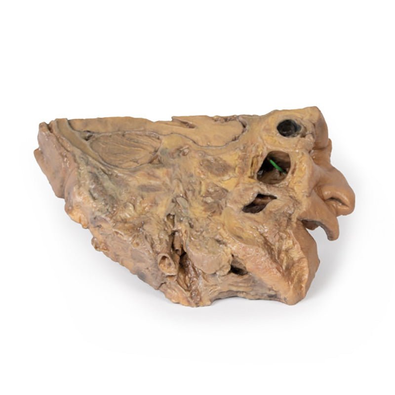

3D Printed Sinus Pathways
Item # MP1106
$1,164.00
$1,295.00
You save $131.00
Need an estimate?
Click Add To Quote

Features & Specifications
-
 by
by
A trusted GT partner -
FREE Shipping
U.S. Contiguous States Only -
 3D Printed Model
from a real specimen -
 Gov't pricing
Available upon request
 by
by 
Frequently Bought Together

3D Printed Sinus Pathways
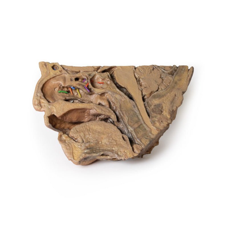
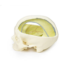
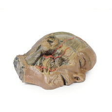
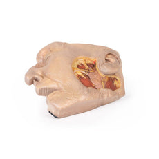
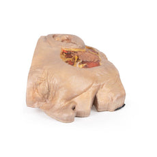
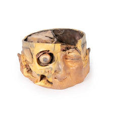
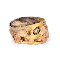
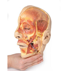
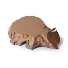
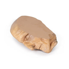
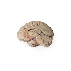
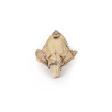
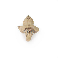
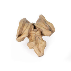
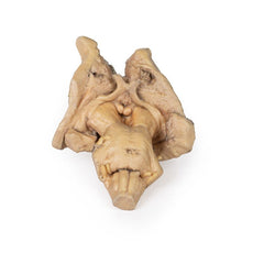
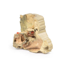
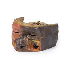
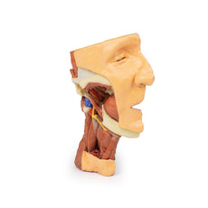
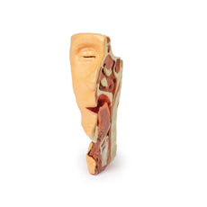
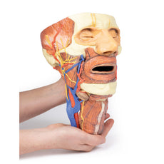
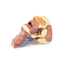
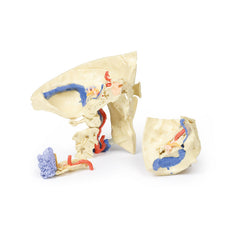
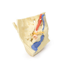
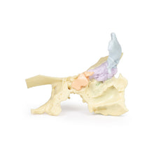
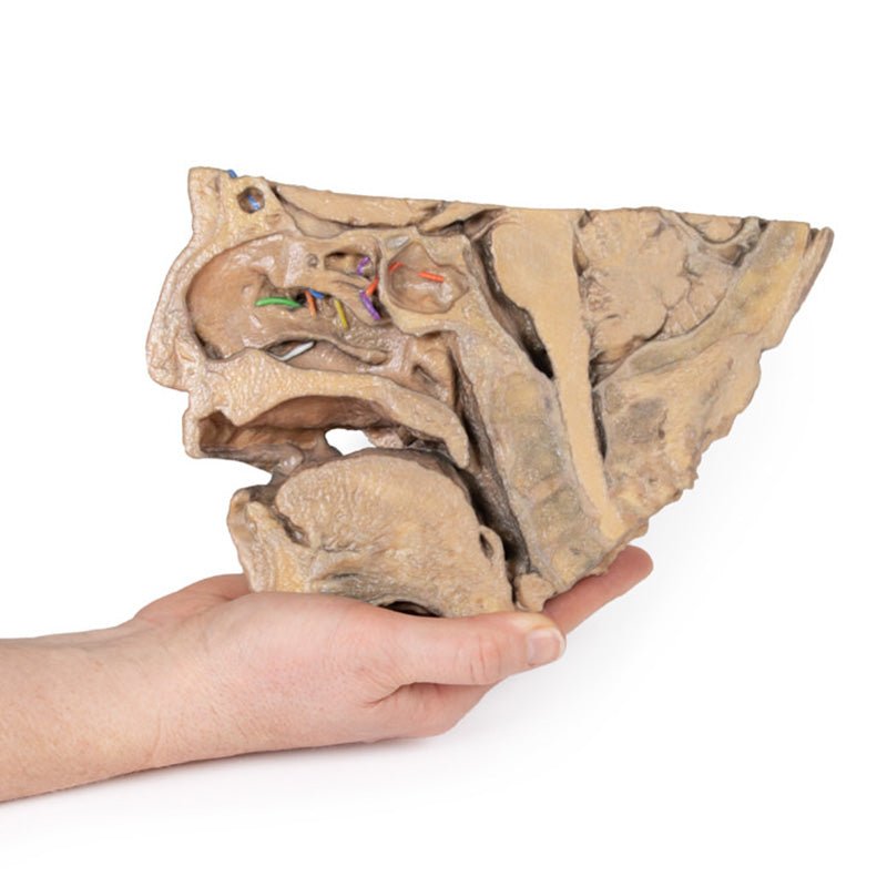
This 3D model provides a midsagittal to parasagittal segment of a right head to demonstrate the relationships and
passageways of the paranasal sinuses. These passageways have been highlighted with thin coloured markers to indicate
the relationship of these communicating routes between the paranasal sinuses and the nasal cavity.
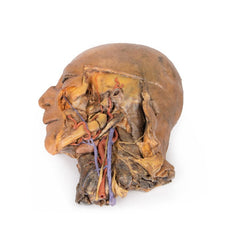
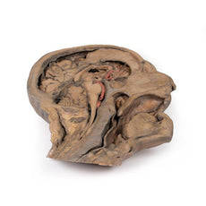
Starting
anteriorly in the nasal cavity, the opening of the nasolacrimal duct (white) is present just deep to the inferior
nasal conchae. The middle nasal concha has been sectioned to allow for a clear view of the opening of the maxillary
sinus (visible in the parasagittal plane) across the semilunar hiatus (green), as well as the drainage of the
frontal sinus (blue; with the sinus visible superiorly in the section and in the transverse cut through the
specimen) and the anterior (orange) and middle (yellow) ethmoidal cells. The opening of the posterior ethmoidal
cells into the superior meatus is shown through the purple marker, which is visible within a small opened window
into the ethmoid just superior to the nasal cavity. Finally, the opening of the sphenoid sinus is marked in red and
visible through the opened sphenoid sinus itself just superior to the nasopharyngeal region.
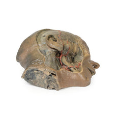
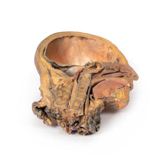
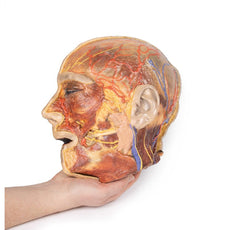
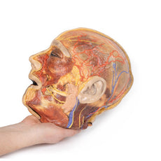
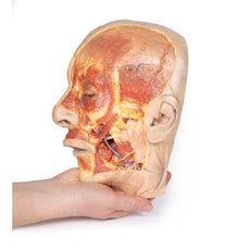
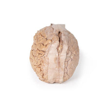
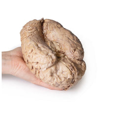
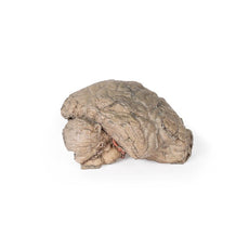
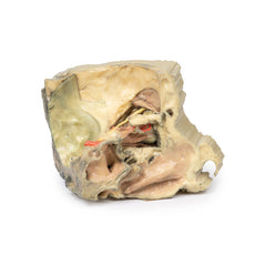
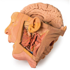
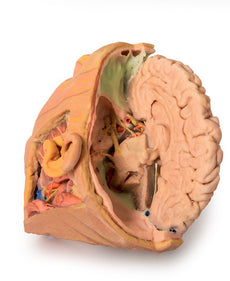
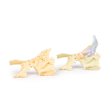
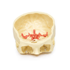
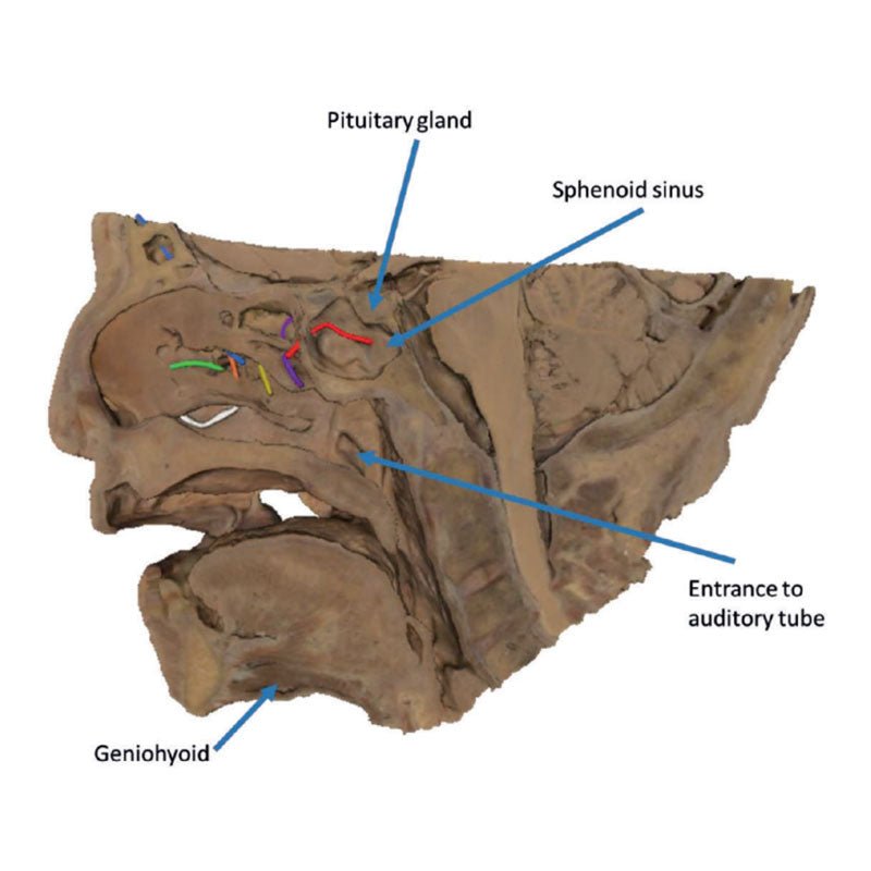
In addition to these
pathways, this 3D model also captures some of the surrounding anatomy within the section. Visible in the midsagittal
view are the other primary structures of the nasal cavity from the nostril to the opening of the auditory tube
posteriorly. The soft palate and uvula are preserved, as is the rest of the pharynx just to the level of the
epiglottis and collapsed laryngeal region at the inferior part of the preserved specimen. The oral cavity is
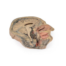
displayed in cross section, with distinct genioglossus and geniohyoid muscles. In the cranial cavity, parts of the
brain are preserved including the inferior parts of the frontal lobe and the right optic nerve/chiasm/tract. The
pituitary gland is visible in cross-section just superior to the sphenoid sinus. The pons, medulla oblongata, and
most of the cerebellum are present, with a small part of the tentorium cerebelli separating the cerebellum from the
right occipital lobe of the cerebrum. On the parasagittal side of the specimen there is a continuation of the
tentorium cerebelli separating these parts of the brain, with clear cross-sections of the transverse sinus and part
of the sigmoid sinus on either side of the cerebellum. Overlying this is a small part of the medial temporal lobe of
the cerebellum with part of the anterior horn of the lateral ventricle deep within the lobe.
Handling Guidelines for 3D Printed Models
GTSimulators by Global Technologies
Erler Zimmer Authorized Dealer
These items normal warranty are two years, however the warranty doesn’t cover “wear and tear”. The manufacturer does have 100% quality control on these models.
The models are very detailed and delicate. With normal production machines you cannot realize such details like shown in these models.
The printer used is a color-plastic printer. This is the most suitable printer for these models.
The plastic material is already the best and most suitable material for these prints. (The other option would be a kind of gypsum, but this is way more fragile. You even cannot get them out of the printer without breaking them).The huge advantage of the prints is that they are very realistic as the data is coming from real human specimen. Nothing is shaped or stylized.
The users have to handle these prints with utmost care. They are not made for touching or bending any thin nerves, arteries, vessels etc. The 3D printed models should sit on a table and just rotated at the table.
Related Products


$2,784.00
$3,095.00
Free shipping
3D Printed Sagittal Section of Head with Infratemporal Fossa Dissection
Item # MP1104

$1,132.00
$1,259.00
Free shipping
3D Printed Parotid Gland and Facial Nerve Dissection
Item # MP1112

$2,914.00
$3,239.00
Free shipping
3D Printed Sagittal Section of Head and Neck with Infratemporal Fossa and Carotid Sheath Dissection
Item # MP1111


$2,329.00
$2,589.00
Free shipping
3D Printed Superficial Facial Nerves & Parotid Gland
Item # MP1109


$2,799.00
$3,111.00
Free shipping
3D Printed Parasagittal Section of the Head and Neck
Item # MP1107

$2,185.00
$2,429.00
Free shipping
3D Printed Median Section Through Head Sagittal Section of Head with Deep Dissection
Item # MP1105


$450.00
$501.00
3D Printed Brain Stem, Isolated Anatomy From Midbrain to Medulla Oblongata
Item # MP1101


$8,529.00
$9,374.00
Free shipping
3D Printed Head, Neck, Shoulder and Thorax Replica with Angiosomes
Item # MP1250
by — Item # MP1106
3D Printed Sinus Pathways
$1,164.00
$1,295.00
Add to Cart
Add to Quote
