

Your shopping cart is empty.
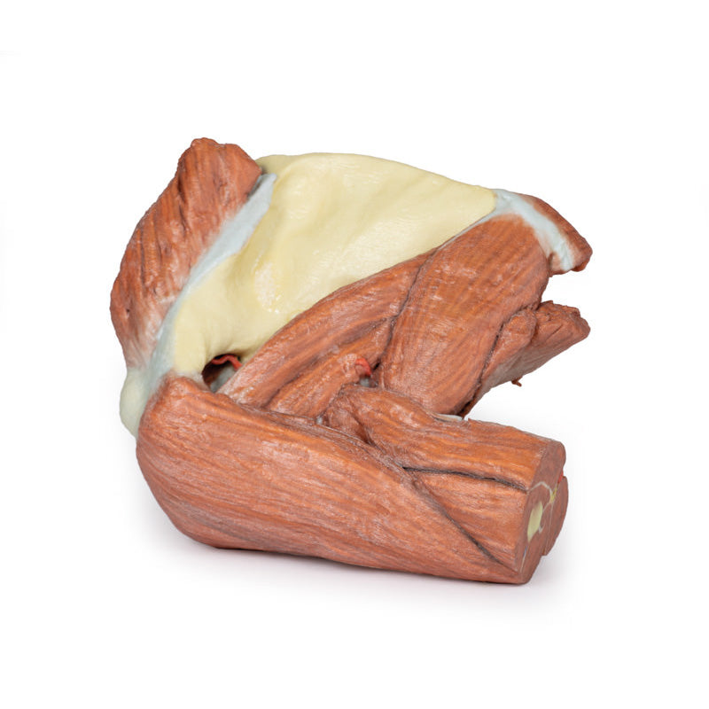
3D Printed Left Shoulder Superficial muscles and brachial artery
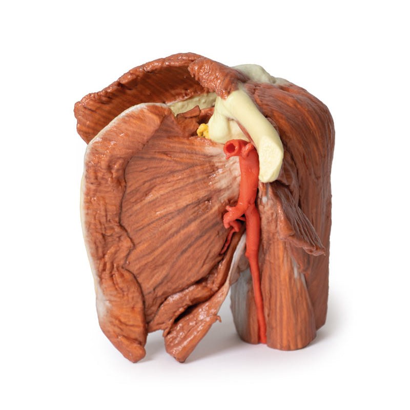
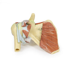
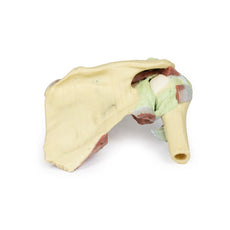
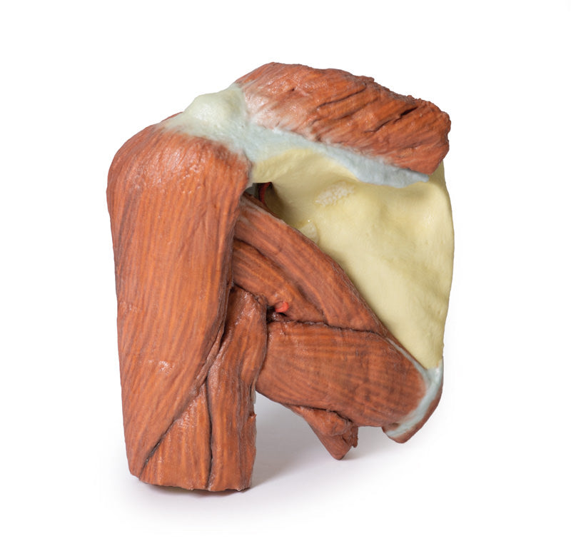
The muscles attached to the clavicle have been preserved including the subclavius muscle attachment to the inferior border of the clavicle and the deltoid covering the lateral aspect of the proximal upper limb (overlying the origins of the long head of biceps brachii and the lateral head of triceps brachii). The clavicular head of the pectoralis major has been preserved. On the posterior aspect the superior fibers of trapezius can also be observed where they attach attached to the posterior border of the lateral third of the clavicle, and to the acromion process and the spine of the scapula. Other muscles attached to the scapula which have been preserved include the subscapularis and serratus anterior on the anterior or costal aspect. Inspection of the anterior aspect reveals that the pectoralis minor insertion onto the coracoid process of the scapula has been preserved. Posteriorly the teres major and teres minor muscles are clearly visible arising from the lateral border of the scapula.
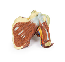
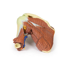
Supraspinatus is preserved but infraspinatus has partly been removed to show branches of the suprascapular artery passing from the supraspinous fossa around the base of the spine to enter the infraspinous fossa housing the infraspinatus muscle. A small part of the omohyoid attachment is also visible above the suprascapular ligament.
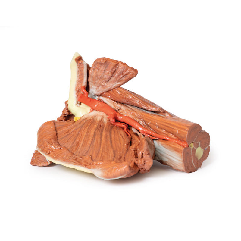
The axillary artery below the inferior border of the clavicle can be seen to give off the thoracoacromial branch anteriorly and just slightly more distally the suprascapular artery can be seen passing posteriorly. Coursing distally, it gives off posterior branches of the circumflex scapular and subscapular arteries. The anterior and posterior circumflex humeral arteries are hidden from view when viewed from in front, however the latter artery can be seen deep to the posterior fibres of deltoid as it emerges though quadrangular space. Below the inferior border of teres major the axillary artery becomes the brachial artery. The radial collateral artery is visible arising from the brachial artery. The axillary artery becomes the brachial artery beyond the lower margin of the teres major muscle.
The muscles of the proximal upper limb have all been preserved, and those of the superficial layer, i.e. long head of biceps brachii, and long and lateral heads of triceps brachii, can be observed to form a complete layer of musculature around the humerus. The cross section of the mid shaft of the humerus nicely displays the relations of the major neurovascular bundles and the muscles in the anterior and posterior compartments. A small remnant of the suprascapular nerve passing under the suprascapular ligament is visible.
GTSimulators by Global Technologies
Erler Zimmer Authorized Dealer


3D Printed Left Shoulder Superficial muscles and brachial artery
Item # MP1523
$1,722.00
$1,935.00
You save $213.00
Need an estimate?
Click Add To Quote

Features & Specifications
-
 by
by
A trusted GT partner -
FREE Shipping
U.S. Contiguous States Only -
 3D Printed Model
from a real specimen -
 Gov't pricing
Available upon request
 by
by 
Frequently Bought Together
3D Printed Left Shoulder Superficial muscles and axillary/brachial artery
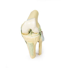
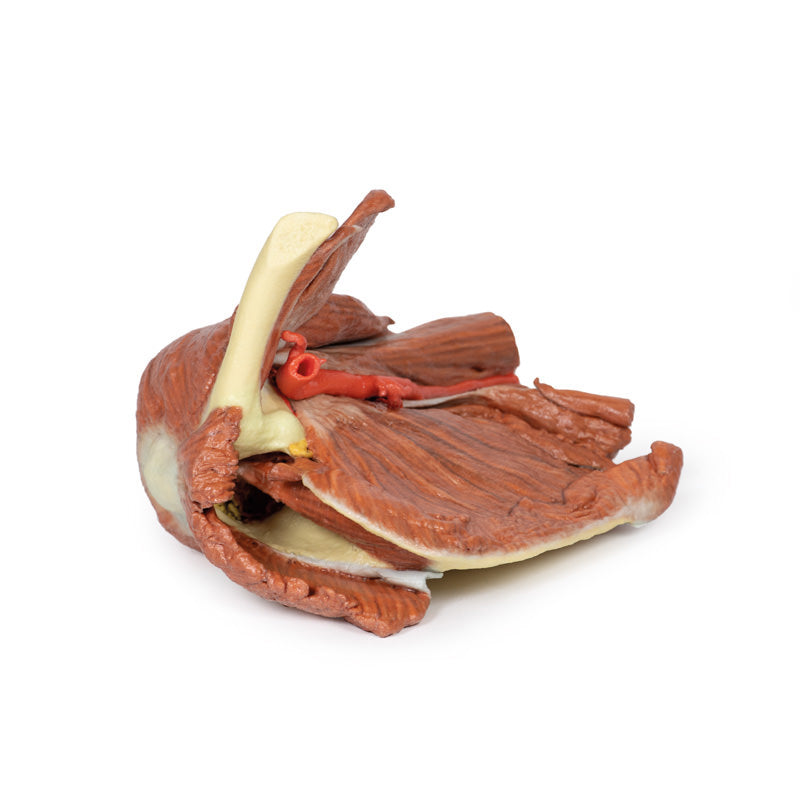
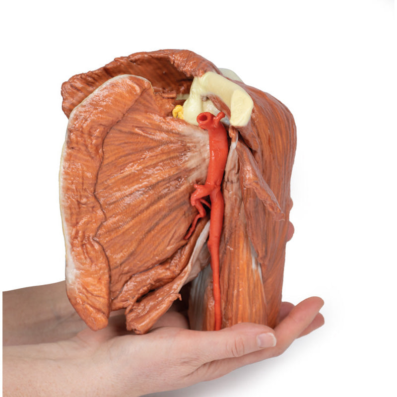
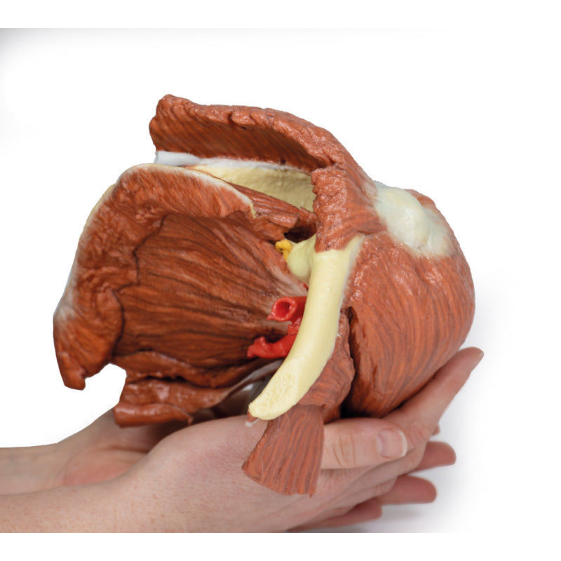
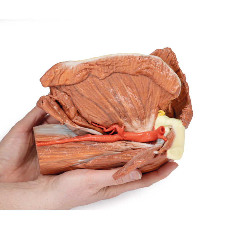
This printed 3D left shoulder specimen consists of the scapula, humerus (sectioned near midshaft) and clavicle (sectioned at midshaft) with the superficial muscles around the shoulder joint, the rotator cuff muscles and the axillary artery as it progresses distally to become the brachial artery.The muscles attached to the clavicle have been preserved including the subclavius muscle attachment to the inferior border of the clavicle and the deltoid covering the lateral aspect of the proximal upper limb (overlying the origins of the long head of biceps brachii and the lateral head of triceps brachii). The clavicular head of the pectoralis major has been preserved. On the posterior aspect the superior fibers of trapezius can also be observed where they attach attached to the posterior border of the lateral third of the clavicle, and to the acromion process and the spine of the scapula. Other muscles attached to the scapula which have been preserved include the subscapularis and serratus anterior on the anterior or costal aspect. Inspection of the anterior aspect reveals that the pectoralis minor insertion onto the coracoid process of the scapula has been preserved. Posteriorly the teres major and teres minor muscles are clearly visible arising from the lateral border of the scapula.
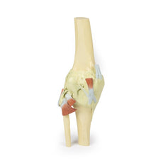
Supraspinatus is preserved but infraspinatus has partly been removed to show branches of the suprascapular artery passing from the supraspinous fossa around the base of the spine to enter the infraspinous fossa housing the infraspinatus muscle. A small part of the omohyoid attachment is also visible above the suprascapular ligament.
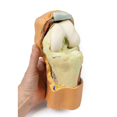
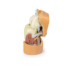
The axillary artery below the inferior border of the clavicle can be seen to give off the thoracoacromial branch anteriorly and just slightly more distally the suprascapular artery can be seen passing posteriorly. Coursing distally, it gives off posterior branches of the circumflex scapular and subscapular arteries. The anterior and posterior circumflex humeral arteries are hidden from view when viewed from in front, however the latter artery can be seen deep to the posterior fibres of deltoid as it emerges though quadrangular space. Below the inferior border of teres major the axillary artery becomes the brachial artery. The radial collateral artery is visible arising from the brachial artery. The axillary artery becomes the brachial artery beyond the lower margin of the teres major muscle.
The muscles of the proximal upper limb have all been preserved, and those of the superficial layer, i.e. long head of biceps brachii, and long and lateral heads of triceps brachii, can be observed to form a complete layer of musculature around the humerus. The cross section of the mid shaft of the humerus nicely displays the relations of the major neurovascular bundles and the muscles in the anterior and posterior compartments. A small remnant of the suprascapular nerve passing under the suprascapular ligament is visible.
Download:

GTSimulators by Global Technologies
Erler Zimmer Authorized Dealer
These items normal warranty are two years, however the warranty doesn’t cover “wear and tear”. The manufacturer does have 100% quality control on these models.
The models are very detailed and delicate. With normal production machines you cannot realize such details like shown in these models.
The printer used is a color-plastic printer. This is the most suitable printer for these models.
The plastic material is already the best and most suitable material for these prints. (The other option would be a kind of gypsum, but this is way more fragile. You even cannot get them out of the printer without breaking them).The huge advantage of the prints is that they are very realistic as the data is coming from real human specimen. Nothing is shaped or stylized.
The users have to handle these prints with utmost care. They are not made for touching or bending any thin nerves, arteries, vessels etc. The 3D printed models should sit on a table and just rotated at the table.
Related Products

$1,013.00
$1,138.00
Free shipping
3D Printed Shoulder with deep dissection of the left shoulder
Item # MP1525


$1,199.00
$1,358.00
Free shipping
3D Printed Shoulder with deep dissection of a right shoulder
Item # MP1527
by — Item # MP1523
3D Printed Left Shoulder Superficial muscles and brachial artery
$1,722.00
$1,935.00
Add to Cart
Add to Quote
