

Your shopping cart is empty.
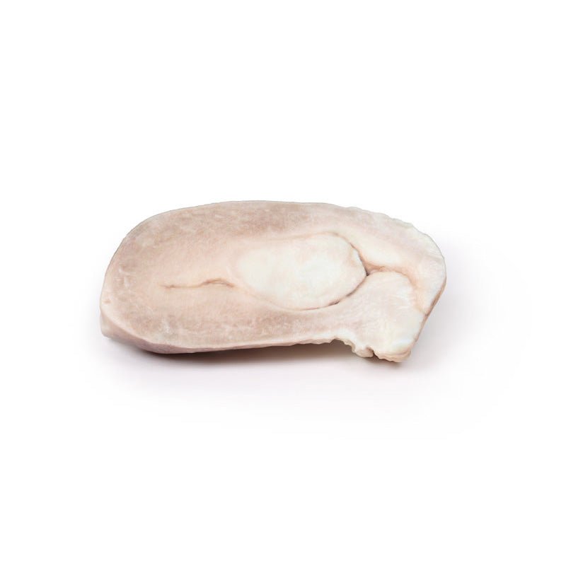
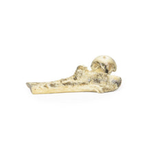
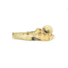
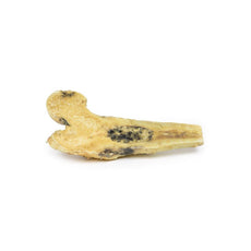
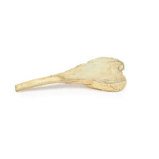
3D Printed Uterine Leiomyoma
 Handling Guidelines for 3D Printed Models
GTSimulators by Global Technologies
Erler Zimmer Authorized Dealer
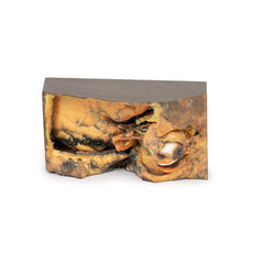
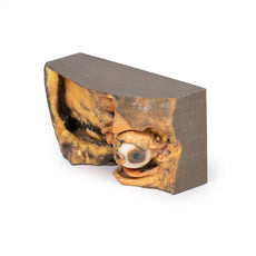
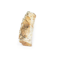
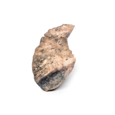
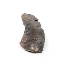
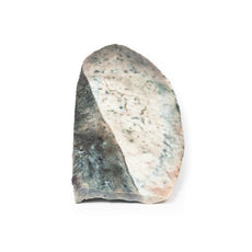
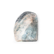
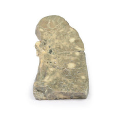
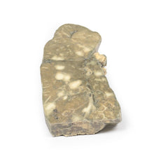
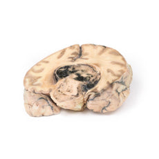
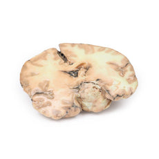

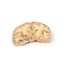
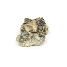
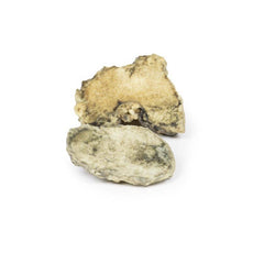
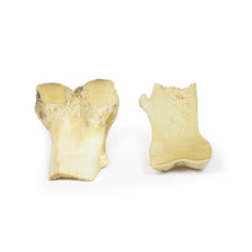

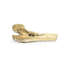
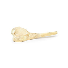
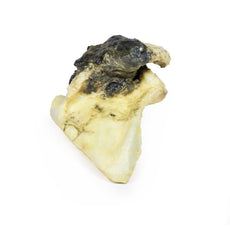
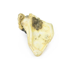
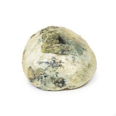
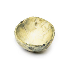
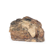
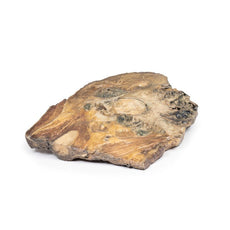
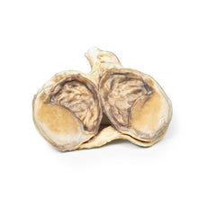
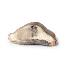


3D Printed Uterine Leiomyoma
Item # MP2107
$401.00
$447.00
You save $46.00
Need an estimate?
Click Add To Quote

Features & Specifications
-
 by
by
A trusted GT partner -
 3D Printed Model
from a real specimen -
 Gov't pricing
Available upon request
 by
by 
Frequently Bought Together
3D Printed Uterine Leiomyoma
Clinical History
A 30-year old female presents with inability to conceive. She also reports a
history of intermittent pelvic discomfort, menorrhagia and painful periods. On examination a pelvic mass was
palpable. All of her blood tests were within normal ranges. A pelvic ultrasound showed a hypoechoic mass within the
myometrium of her uterus. She went for hysteroscopic myomectomy but unfortunately complications meant her surgery
was converted to an emergency hysterectomy. She made a full recovery post-op.
Pathology
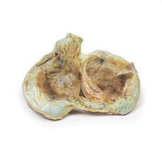
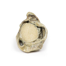
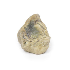
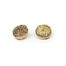
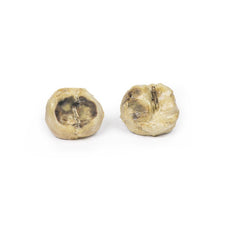
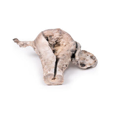
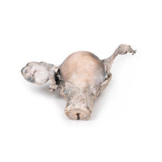
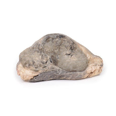
The specimen includes the cervix, body and fundus of the uterus. The uterus, which is
of normal size, has been cut in the sagittal plane. A large ovoid mass approximately 4cm x 2cm protrudes into the
uterine cavity and extends as far inferiorly as the opening of the cervix. It originates from the posterior aspect
of the uterus. The cervical canal is clearly visible.
Further Information
Uterine leiomyomas , also called fibroids, are the most common pelvic tumours
in females. They are present in almost 25% of reproductive females. They are benign tumours arising from smooth
muscle and fibroblasts of the myometrium. They usually involve the myometrium of the uterine body. Rarely they can
affect the lower uterus or cervix. Leiomyomas can occur as single lesions or multiple and can grow very large. There
are rare variants, which can extend and spread distally but are still considered benign: e.g. the benign
metastasizing leiomyoma, which commonly spread to the lining; or disseminated peritoneal leiomyomatosis, which
appears on the peritoneum covering the uterus
Risk factors for developing fibroid include being of reproductive
age, being a black woman and early menarche. Higher parity has been found to be protective. Most leiomyomas have
normal karyotypes but there are some which show mutation in the HMG genes. Transformation into malignant
leiomyosarcoma is very rare.
Common symptoms of uterine fibroids include abnormal vaginal bleeding, pelvic pain,
dyspareunia, dysmenorrhea and symptoms of pelvic structure compression such as urinary symptoms or venous
compression symptoms. Leiomyomas can decrease fertility and in pregnant females increase the rate of early pregnancy
loss, fetal malpresentation and postpartum hemorrhage. Pelvic ultrasound is usually used to diagnose leiomyomas. CT
and MRI scans are rarely used to diagnose.
Leiomyomas can grow but can also regress. Treatment is reserved for persistently or severely symptomatic fibroids. Hormonal treatment may be used to regulate irregular menstrual bleeding symptoms. Surgical treatments include myomectomy (removal of fibroids from myomectomy), hysterectomy, myolysis (thermal ablation of leiomyoma) and uterine artery ablation/embolisation.
Download:
Handling Guidelines for 3D Printed Models
GTSimulators by Global Technologies
Erler Zimmer Authorized Dealer
These items normal warranty are two years, however the warranty doesn’t cover “wear and tear”. The manufacturer does have 100% quality control on these models.
The models are very detailed and delicate. With normal production machines you cannot realize such details like shown in these models.
The printer used is a color-plastic printer. This is the most suitable printer for these models.
The plastic material is already the best and most suitable material for these prints. (The other option would be a kind of gypsum, but this is way more fragile. You even cannot get them out of the printer without breaking them).The huge advantage of the prints is that they are very realistic as the data is coming from real human specimen. Nothing is shaped or stylized.
The users have to handle these prints with utmost care. They are not made for touching or bending any thin nerves, arteries, vessels etc. The 3D printed models should sit on a table and just rotated at the table.
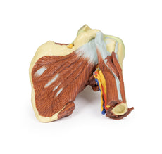
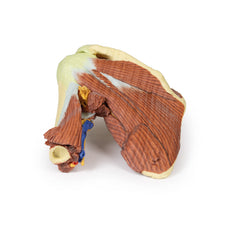
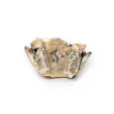
Related Products

$1,013.00
$1,138.00
Free shipping
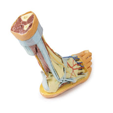
3D Printed Shoulder with deep dissection of the left shoulder
Item # MP1525


by — Item # MP2107
3D Printed Uterine Leiomyoma
$401.00
$447.00
Add to Cart
Add to Quote
