

Your shopping cart is empty.
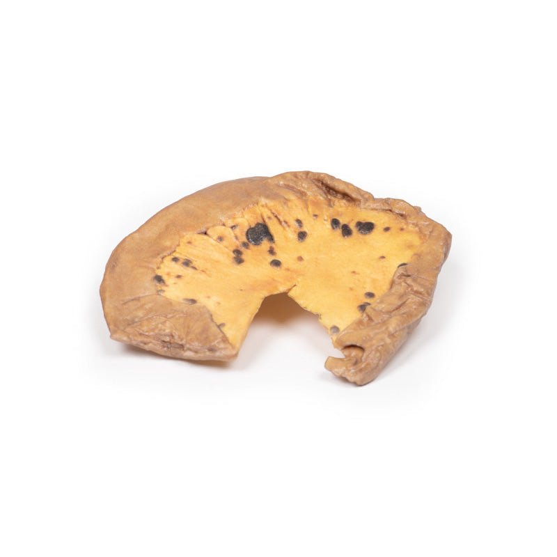
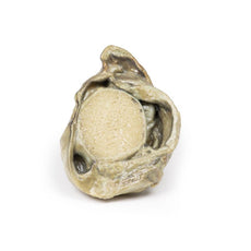
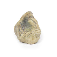
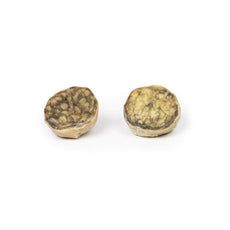
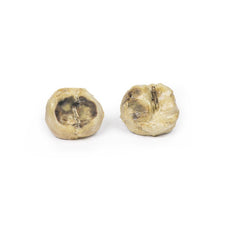
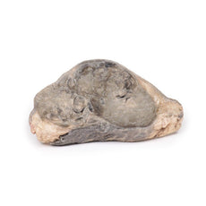
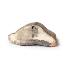
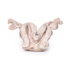
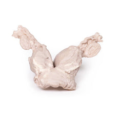
3D Printed Mesenteric Metastases from Malignant Melanoma
 Handling Guidelines for 3D Printed Models
GTSimulators by Global Technologies
Erler Zimmer Authorized Dealer
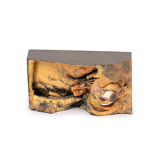
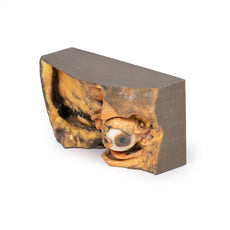
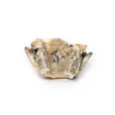
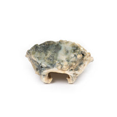
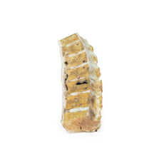
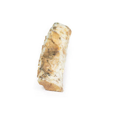
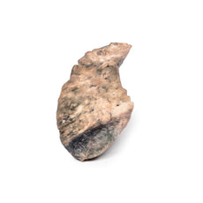
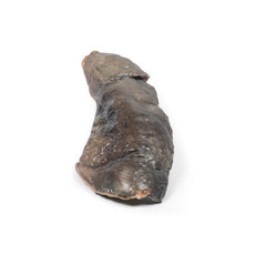
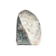
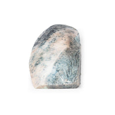
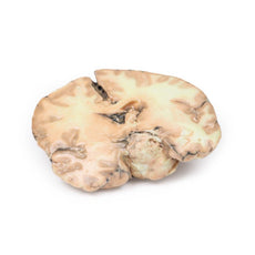


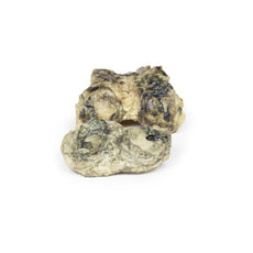
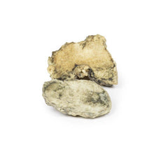
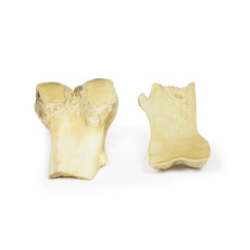

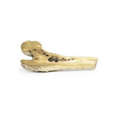
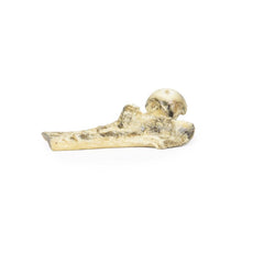
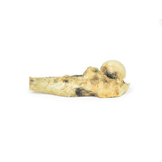
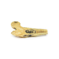
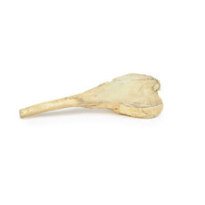
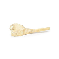
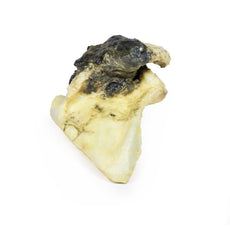
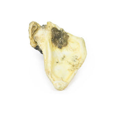
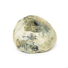
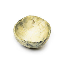
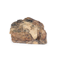
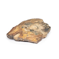
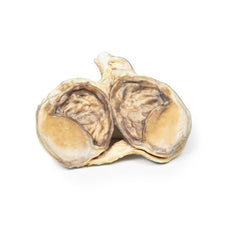
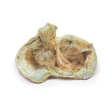


3D Printed Mesenteric Metastases from Malignant Melanoma
Item # MP2083
$273.00
$305.00
You save $32.00
Need an estimate?
Click Add To Quote

Features & Specifications
-
 by
by
A trusted GT partner -
 3D Printed Model
from a real specimen -
 Gov't pricing
Available upon request
 by
by 
Frequently Bought Together
3D Printed Mesenteric Metastases from Malignant Melanoma
Clinical History
A 44-year-old man had a skin lesion on his back that grew slowly. At
presentation at A&E several years later, he complained of bone pain, and had hepatomegaly and a pleural
effusion. He died shortly afterwards.
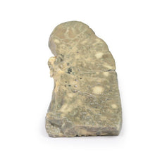
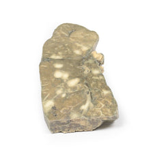
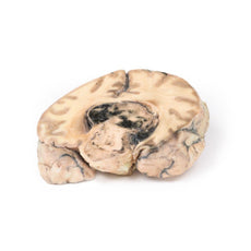
Pathology
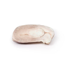
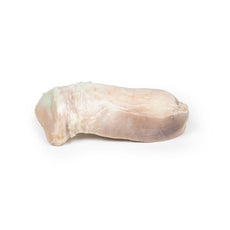
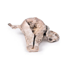
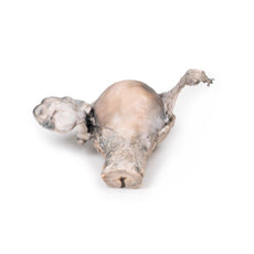
The specimen is a loop of small intestine mounted to display the mesentery, which
contains numerous small dark brown, circumscribed nodules varying from pin head size to approximately 1 cm in
diameter. Histology confirmed the diagnosis of metastatic melanoma.
Further Information
The most common form of melanoma is cutaneous melanoma, which develops from
the pigment-producing cells known as melanocytes. In women, they most commonly occur on the legs, while in men they
most commonly occur on the back. About 25% of melanomas develop from moles. Changes in a mole that can indicate
melanoma include an increase in size, irregular edges, change in colour, itchiness or skin ulceration.
Skin melanoma is associated with exposure to UV radiation in sunlight or tanning beds. Other risk factors for
developing melanoma include fair complexion, presence of large number of melanocytic naevi (moles), severe sunburn
as a child, and immunosuppression. It accounts for around 5% of all skin cancer diagnosis but has the highest
mortality rate of all skin cancers. Melanomas typically occur in sun exposed areas as a pigmented lesion with
irregular borders, variegated colour, an asymmetrical shape and which evolves with time.
There are multiple
mutations common in melanoma. Loss of cell cycle control gene from mutation in CDKN2A gene. Mutations in pro-growth
signalling pathways, such as BRAF and PI3K mutations, are seen frequently in melanomas as well as mutations that
activate telomerase, such as the TERT gene. Recognition that melanoma antigens activate host immune responses has
led to promising immunotherapy, which enhances host T-cell identifying of these antigens.
The most common sites for metastasis of melanoma are the lungs, liver, brain and bone as well as regional lymph
nodes, and is highly dependent on the site of the primary tumour. Metastatic melanoma involving the gastrointestinal
tract may present with anaemia, overt bleeding, pain, obstruction, or intussusception. The jejunum and ileum are the
most commonly involved sites, followed by the colon, rectum, and stomach. Surgery has usually been reserved for
patients with the above complications.
The probability of metastatic spread from skin melanoma depends on the
stage of the primary tumour, which is based on tumour depth, mitotic activity and ulceration of the skin as well as
node and solid organ involvement. Diagnosis of melanoma is made with excisional biopsy. Investigation for bone
metastasis is done using blood test (raised Alkaline phosphatase, calcium and LDH), and radiological investigations
most commonly X-ray and CT but MRI and PET scans may also be used. Treatment depends on the stage or the tumour as
well as the genetic and immune profiles of the melanoma. Treatment usually involves surgical resection,
chemotherapy, targeted therapies (e.g. BRAF inhibitors), immunotherapy , radiotherapy or more commonly a combination
of treatments.
Handling Guidelines for 3D Printed Models
GTSimulators by Global Technologies
Erler Zimmer Authorized Dealer
These items normal warranty are two years, however the warranty doesn’t cover “wear and tear”. The manufacturer does have 100% quality control on these models.
The models are very detailed and delicate. With normal production machines you cannot realize such details like shown in these models.
The printer used is a color-plastic printer. This is the most suitable printer for these models.
The plastic material is already the best and most suitable material for these prints. (The other option would be a kind of gypsum, but this is way more fragile. You even cannot get them out of the printer without breaking them).The huge advantage of the prints is that they are very realistic as the data is coming from real human specimen. Nothing is shaped or stylized.
The users have to handle these prints with utmost care. They are not made for touching or bending any thin nerves, arteries, vessels etc. The 3D printed models should sit on a table and just rotated at the table.
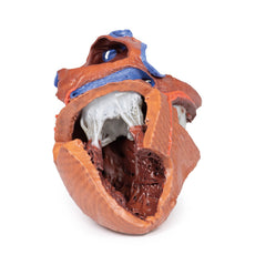
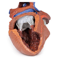
Related Products

$1,013.00
$1,138.00
Free shipping
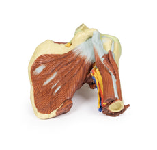
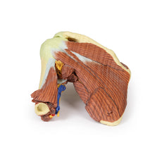
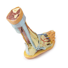
3D Printed Shoulder with deep dissection of the left shoulder
Item # MP1525


by — Item # MP2083
3D Printed Mesenteric Metastases from Malignant Melanoma
$273.00
$305.00
Add to Cart
Add to Quote
