Your shopping cart is empty.
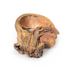
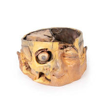
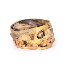
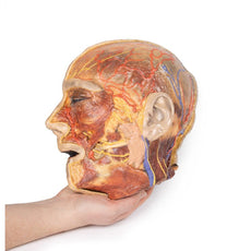
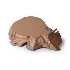
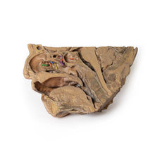
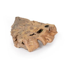
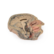
3D Printed Sagittal Section of Head with Infratemporal Fossa Dissection
Item # MP1104Need an estimate?
Click Add To Quote

-
by
A trusted GT partner -
FREE Shipping
U.S. Contiguous States Only -
3D Printed Model
from a real specimen -
Gov't pricing
Available upon request
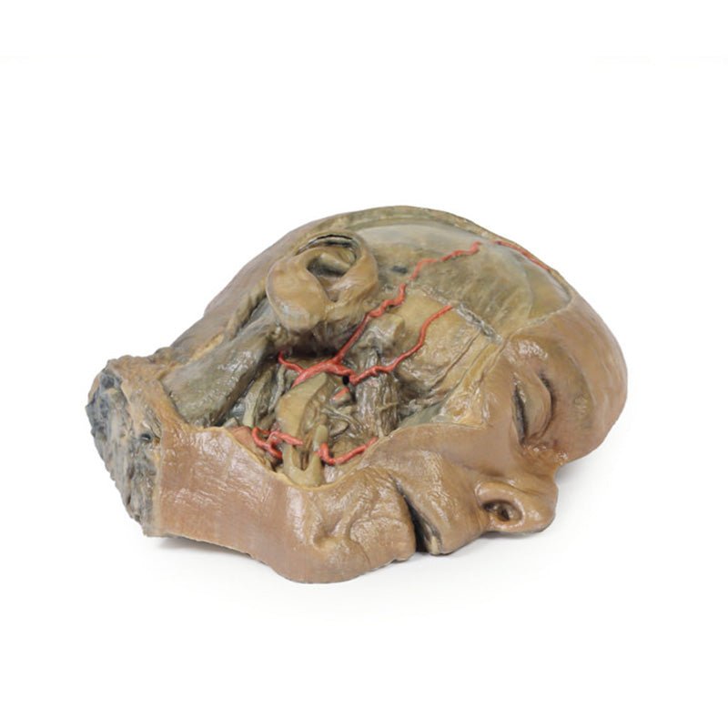
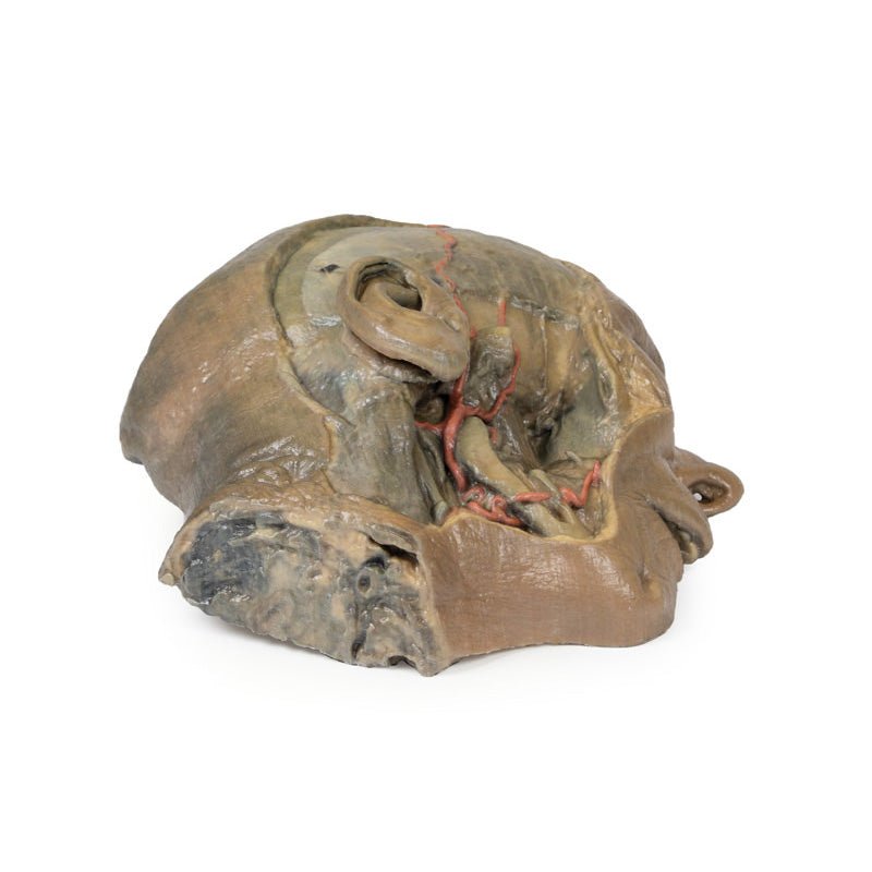
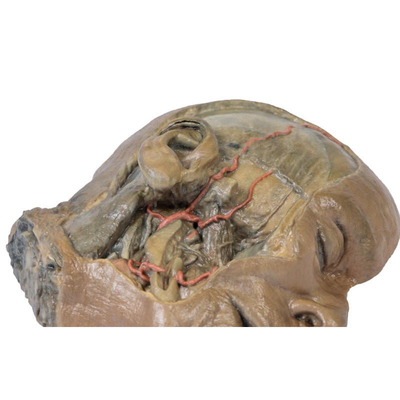
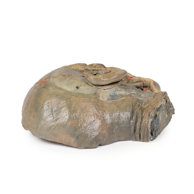
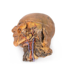
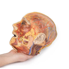
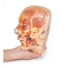
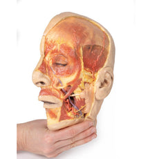
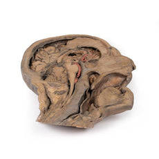
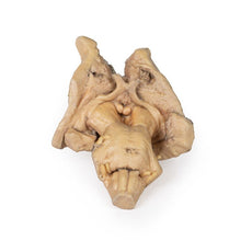
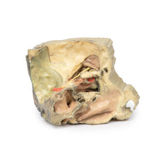
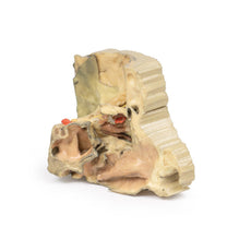
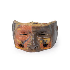
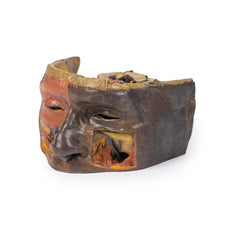
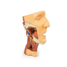
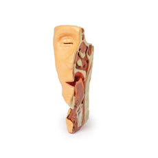
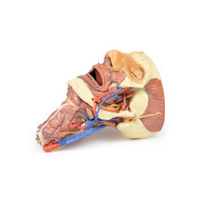
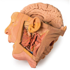
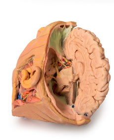
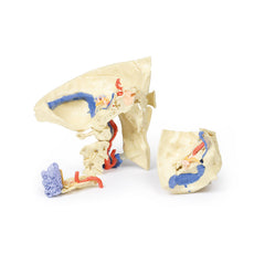
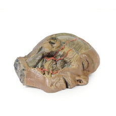
3D Printed Sagittal Section of Head with Infratemporal Fossa Dissection
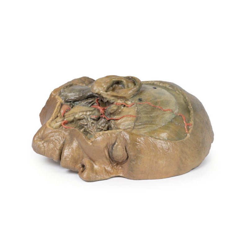
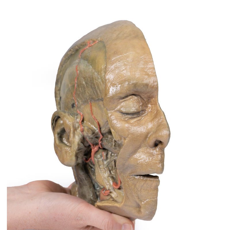
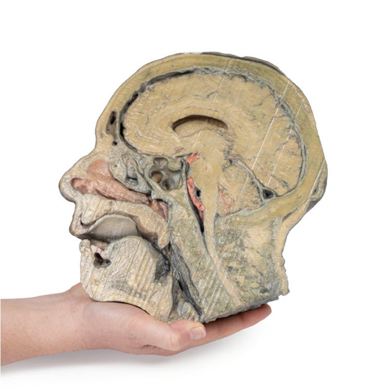
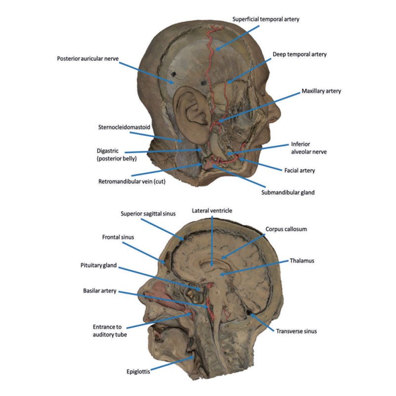
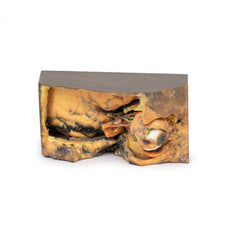
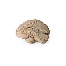
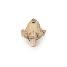
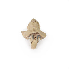
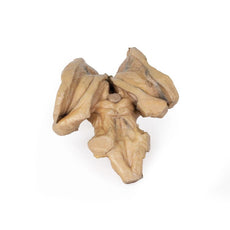
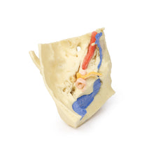
This 3D model provides a combined midsagittal section through the head and
superior neck coupled with a deep dissection into the infratemporal fossa
region and superficial dissection of the scalp.
In the preserved midsagittal section there is preservation of the endocranial
contents, the nasal and oral cavities, and the pharynx to the level of the
laryngeal cartilages. The nasal cavity is preserved nearly intact, except for a
small window excised into the middle nasal concha to expose the ethmoid
air cells. A very large sphenoid sinus exists in the individual just superior
to the torus of the auditory tube in the nasopharynx. The oral cavity and
laryngopharynx are undissected, with the larynx only preserve just distal to the
level of the arytenoid cartilages and not including a clear set of vocal folds.
Within the endocranial cavity, the sectioned brain is slightly off the midagittal
plane, such that neither the superior sagittal sinus nor the third ventricle are
clearly defined - but the lateral ventricle is open and part of the fourth ventricle
is preserved between the pons and cerebellum. The gyri and sulci of the
cerebrum are not well separated, but the cingulate gyrus and corpus callosum
can be separated. Cross-sectioned views of the optic tract, pituitary gland,
superior and inferior colliculi, superior cerebellar peduncle, and transition
between the medulla oblongata and spinal cord are all visible. The tentorium
cerebelli and confluence/transverse sinus is positioned between the
cerebellar hemisphere and occipital lobe. Small portions of the posterior
inferior cerebellar artery, vertebral arteries, basilar artery, and posterior
cerebral and anterior cerebral arteries are visible in section.
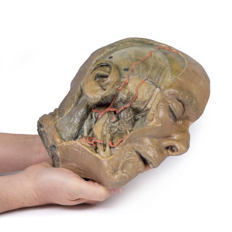
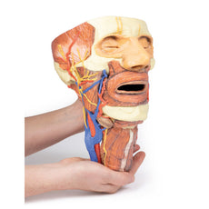
On the opposing side of the model, a superficial and deep dissection has
opened a large window into the anatomy of the lateral scalp and infratemporal
fossa. Across the scalp there is a well preserved posterior auricular nerve
and superficial temporal artery highlighted on the superficial surface of the
temporalis muscle. Anteriorly, the temporalis has been dissected to expose
the deep temporal arteries arising from across the maxillary artery.
The deep level of dissection has exposed parts of the infratemporal fossa
(through partial removal of the mandibular ramus and corpus) and dissection
of retromandibular tissues. At the inferior margin of the dissection window,
the cut edge of the retromandibular vein lies adjacent to the submandibular
gland and the ascending path of the facial artery as it cross towards to angle
of the mouth. Just superior to the cut retromandibular vein is the posterior
belly of the digastric muscle, overlying a small exposure of the deeper
internal jugular vein.
Just posterior to the retained ascending ramus of the mandible are the
external carotid artery and the occipital artery (running in parallel prior
to passing posteriorly). Tracing the external carotid artery superiorly, the
posterior auricular artery, superficial temporal artery, and maxillary artery are
all visible. The maxillary artery passes deep to the lateral pterygoid muscle
and into the infratemporal fossa, reappearing superior to the lateral pterygoid
as it passes into the pterygomaxillary fissure. Along its course, it gives rise
to the posterior deep temporal artery, the inferior alveolar artery (which is
exposed in the dissected mandibular corpus), the anterior deep temporal
artery, and the posterior superior alveolar artery. Finally, the inferior alveolar
nerve can be seen coursing within the opened mandibular corpus, and the
lingual nerve resting on the medial pterygoid. The buccinator muscle is also
retained, with the distal part of the parotid duct preserved as it enters the
muscle towards the oral mucosa
 Handling Guidelines for 3D Printed Models
GTSimulators by Global Technologies
Erler Zimmer Authorized Dealer
The models are very detailed and delicate. With normal production machines you cannot realize such details like shown in these models.
The printer used is a color-plastic printer. This is the most suitable printer for these models.
The plastic material is already the best and most suitable material for these prints. (The other option would be a kind of gypsum, but this is way more fragile. You even cannot get them out of the printer without breaking them).The huge advantage of the prints is that they are very realistic as the data is coming from real human specimen. Nothing is shaped or stylized.
The users have to handle these prints with utmost care. They are not made for touching or bending any thin nerves, arteries, vessels etc. The 3D printed models should sit on a table and just rotated at the table.


