Your shopping cart is empty.
3D Printed Superficial Face

 Handling Guidelines for 3D Printed Models
GTSimulators by Global Technologies
Erler Zimmer Authorized Dealer

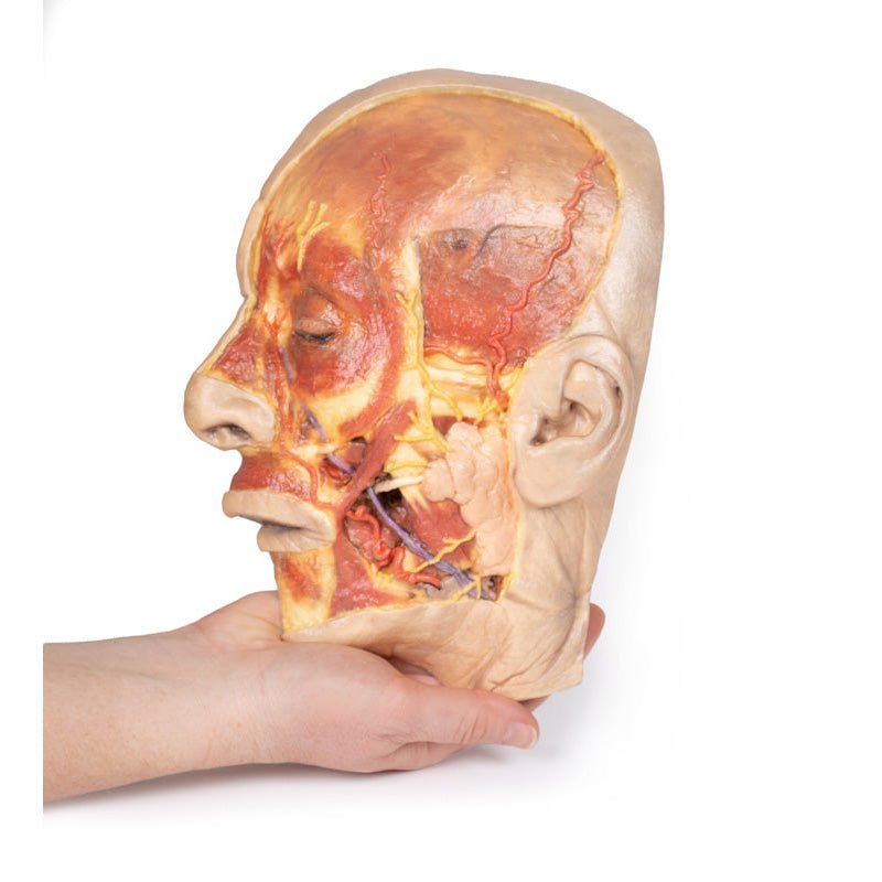

3D Printed Superficial Face
Item # MP1108
$1,778.00
$1,977.00
You save $199.00
Need an estimate?
Click Add To Quote

Features & Specifications
-
 by
by
A trusted GT partner -
FREE Shipping
U.S. Contiguous States Only -
 3D Printed Model
from a real specimen -
 Gov't pricing
Available upon request
 by
by 
Frequently Bought Together
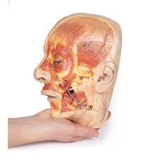
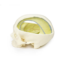
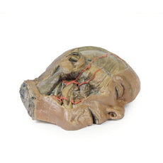
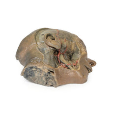
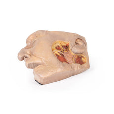
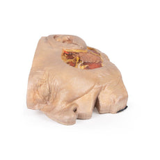
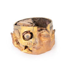
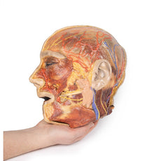
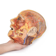
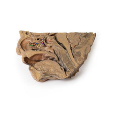
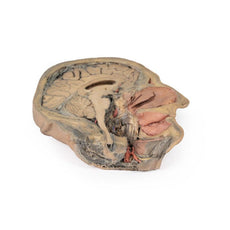
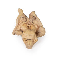
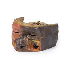
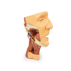
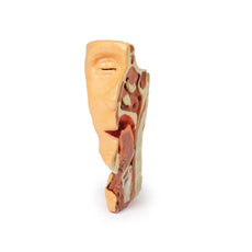
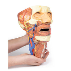
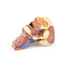
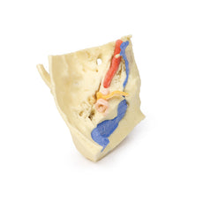
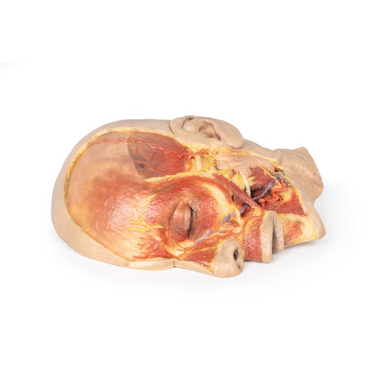
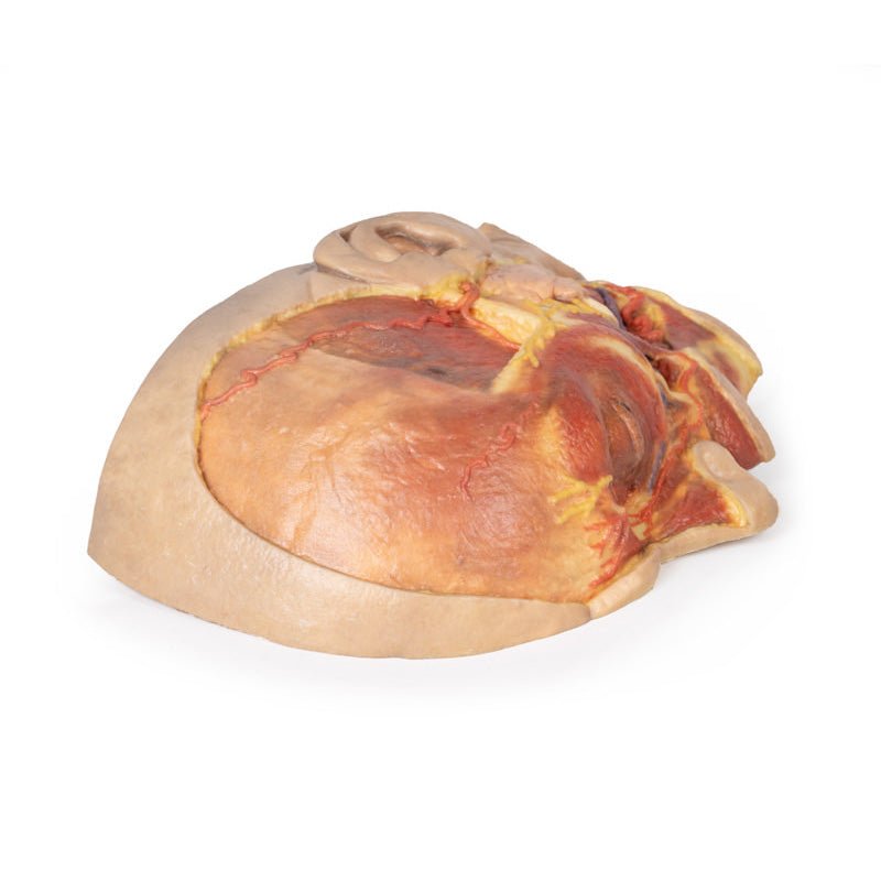
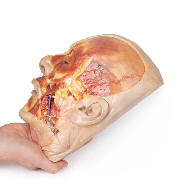
3D Printed Superficial Face
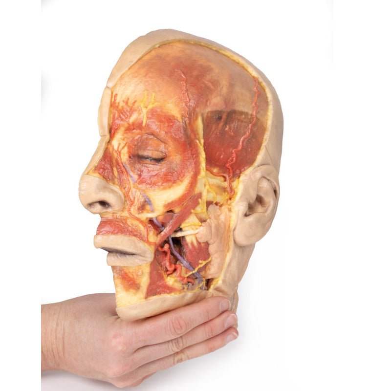
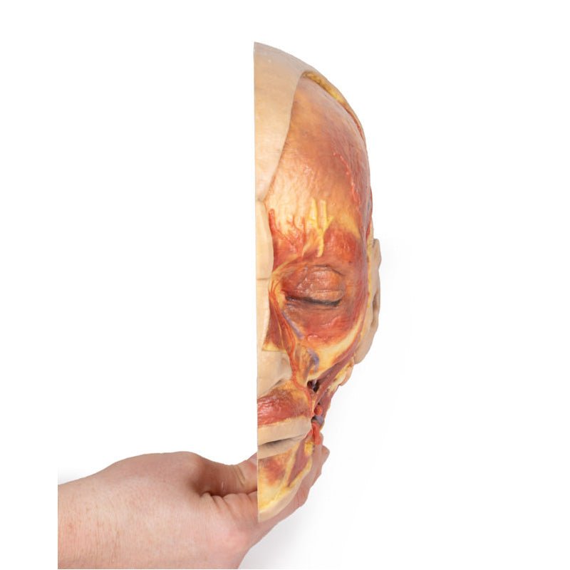
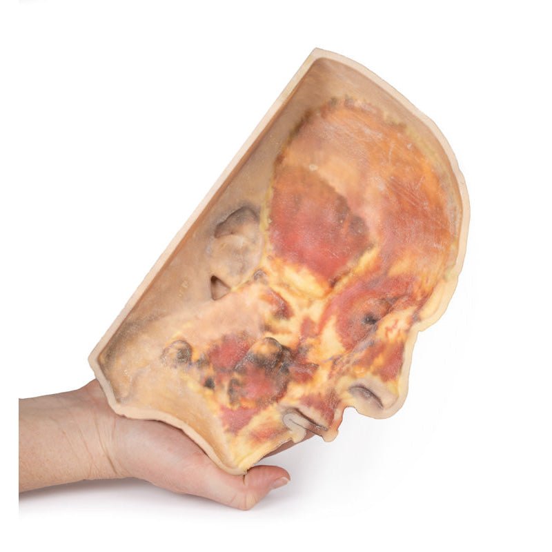
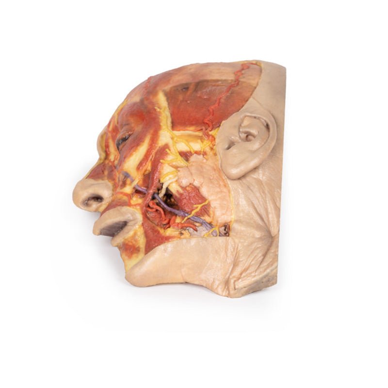
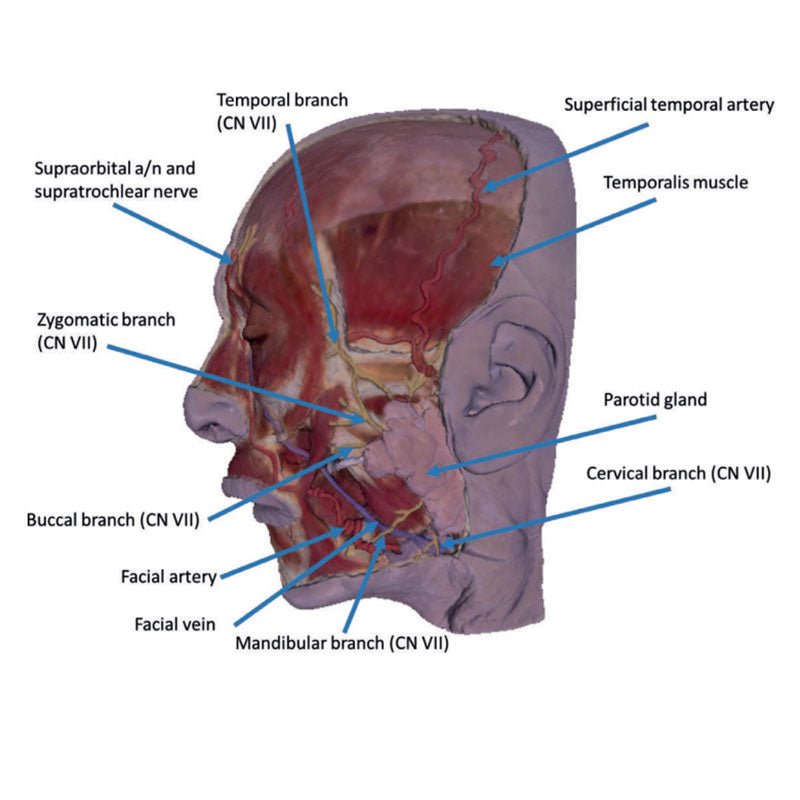
This 3D model presents a superficial dissection of a left face anterior to the ear with false colouring highlighting
a series of neurovascular structures alongside the superficial muscles of facial expression. This compliments the
more expanded superficial dissection of the face and lateral head presented in our HW 45 model. The undissected
regions of the model have been digitally removed.
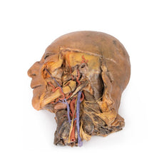
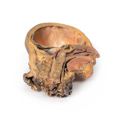
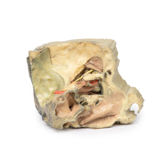
Starting just anterior to the ear, the opened window of
dissection has exposed the parotid gland and associated duct transmitting anterior towards the oral cavity. Exiting
from the margins of the parotid gland are terminal branches of the facial nerve (CN VII), including the cervical,
mandibular, buccal, zygomatic and temporal. The cervical and mandibular branches at the inferior portion of the
dissection window can be seen angling inferiorly and passing superficially relative to the facial vein (which
ascends towards the medial canthus of the eye). The mandibular branch passes just deep to the facial artery, which
runs in parallel with the facial vein. Tracing the pathway of these vessels from the mandible towards the nasal and
orbital regions also provides a checklist of superficial and deep muscles that have been highlighted, from the
masseter deep to the parotid through to the depressor anguli oris, depressor labii inferioris, the zygomaticus major
and minor, the orbicularis oris, the nasalis and levator labii superioris alaeque, the procerus and the orbicularis
oculi.
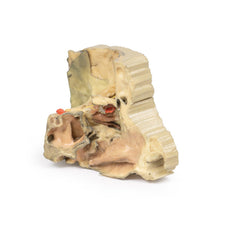
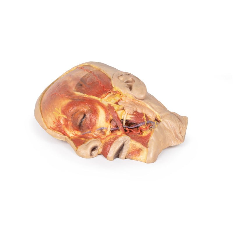
Along the superior margin of the parotid gland the base of the auriculotemporal nerve and the superficial
temporal artery ascends anterior to the ear and rests on a partially dissected temporal fascia to expose part of the
temporalis muscle. Moving anteriorly over the orbit, the supraorbital nerve and supraorbital and supratrochlear
arteries have been highlighted and ascend on the epicranial aponeurosis. Within that layer the deeper frontalis
muscle can be appreciated as a darker shadow within the layer.
Handling Guidelines for 3D Printed Models
GTSimulators by Global Technologies
Erler Zimmer Authorized Dealer
These items normal warranty are two years, however the warranty doesn’t cover “wear and tear”. The manufacturer does have 100% quality control on these models.
The models are very detailed and delicate. With normal production machines you cannot realize such details like shown in these models.
The printer used is a color-plastic printer. This is the most suitable printer for these models.
The plastic material is already the best and most suitable material for these prints. (The other option would be a kind of gypsum, but this is way more fragile. You even cannot get them out of the printer without breaking them).The huge advantage of the prints is that they are very realistic as the data is coming from real human specimen. Nothing is shaped or stylized.
The users have to handle these prints with utmost care. They are not made for touching or bending any thin nerves, arteries, vessels etc. The 3D printed models should sit on a table and just rotated at the table.
Related Products


$2,784.00
$3,095.00
Free shipping
3D Printed Sagittal Section of Head with Infratemporal Fossa Dissection
Item # MP1104

$1,132.00
$1,259.00
Free shipping
3D Printed Parotid Gland and Facial Nerve Dissection
Item # MP1112

$2,914.00
$3,239.00
Free shipping
3D Printed Sagittal Section of Head and Neck with Infratemporal Fossa and Carotid Sheath Dissection
Item # MP1111


$2,329.00
$2,589.00
Free shipping
3D Printed Superficial Facial Nerves & Parotid Gland
Item # MP1109

$2,799.00
$3,111.00
Free shipping
3D Printed Parasagittal Section of the Head and Neck
Item # MP1107


$2,185.00
$2,429.00
Free shipping
3D Printed Median Section Through Head Sagittal Section of Head with Deep Dissection
Item # MP1105


$450.00
$501.00
3D Printed Brain Stem, Isolated Anatomy From Midbrain to Medulla Oblongata
Item # MP1101


$8,529.00
$9,374.00
Free shipping
3D Printed Head, Neck, Shoulder and Thorax Replica with Angiosomes
Item # MP1250
by — Item # MP1108
3D Printed Superficial Face
$1,778.00
$1,977.00
Add to Cart
Add to Quote