

Your shopping cart is empty.
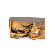
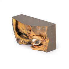
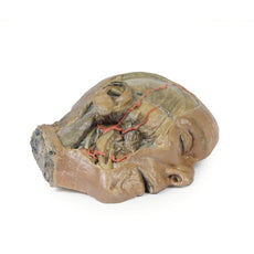
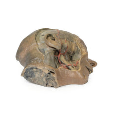
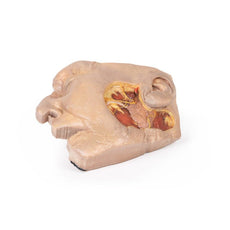
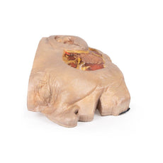
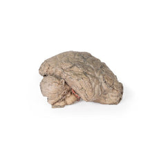
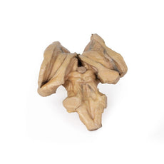
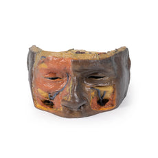
3D Printed Head and Visceral Column of the Neck


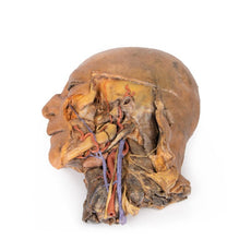
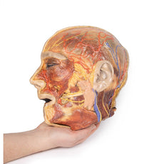
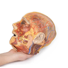
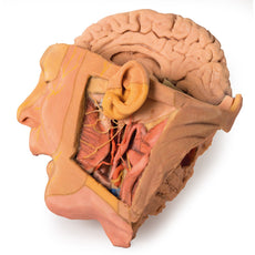
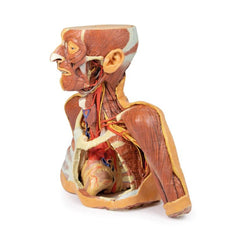
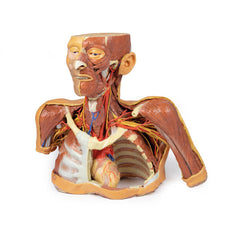
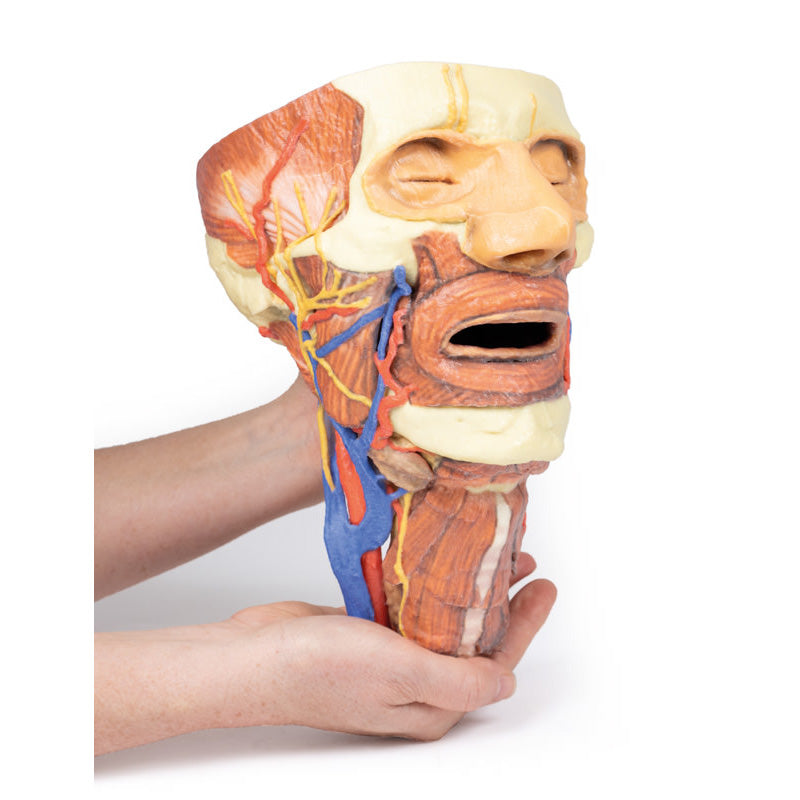
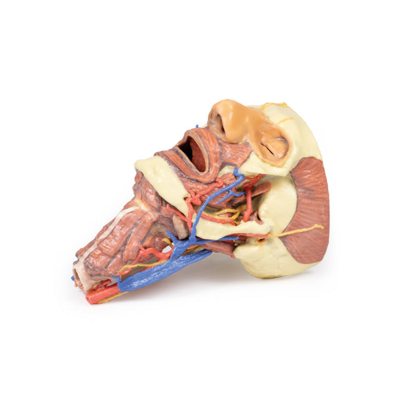
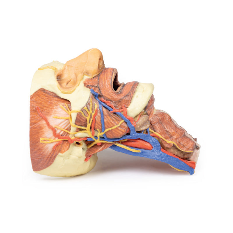
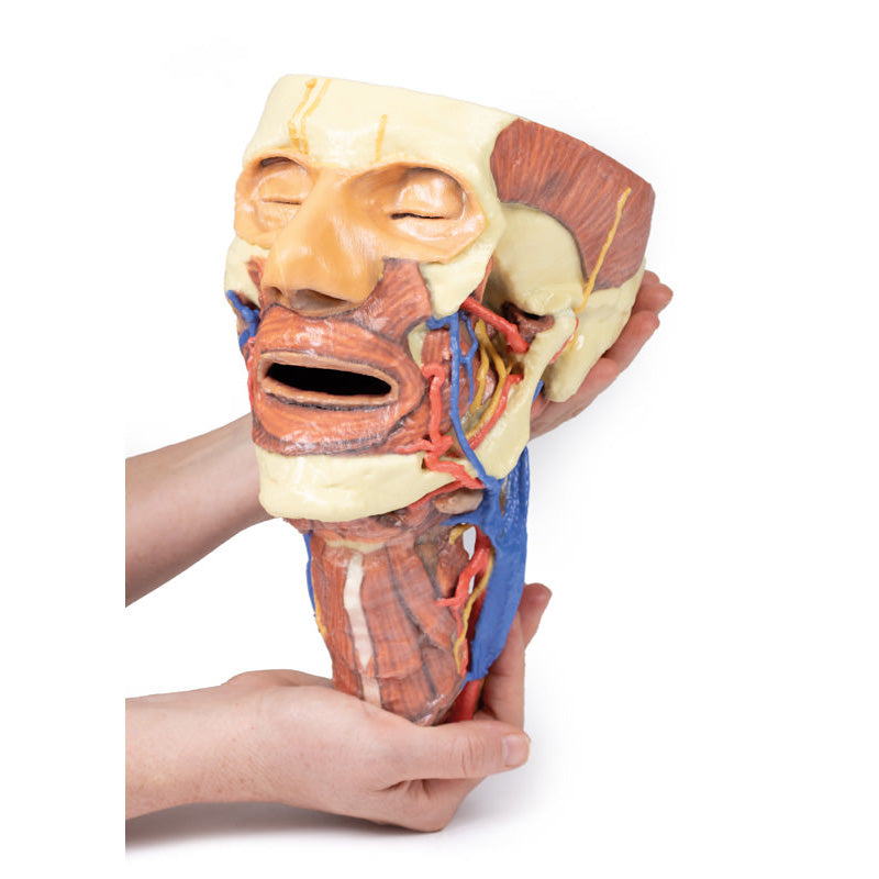
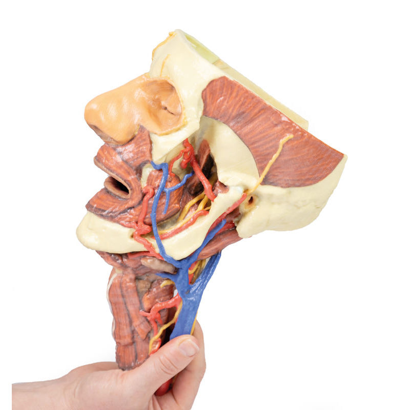
The face: On the right side of the head the parotid gland has been removed to reveal the facial nerve and all its branches (temporal, zygomatic, buccal, marginal mandibular and cervical) and demonstrate the spatial relations of structures embedded in the gland from superficial to deep (facial nerve, retromandibular vein, external carotid artery). In the surrounding region the temporalis, masseter and posterior belly of digastric are exposed, as are and the facial artery, transverse facial artery and superficial temporal artery. The facial vein and transverse facial vein are clearly visible uniting to form the common facial vein which is joined by the retromandibular vein to form the external jugular vein.
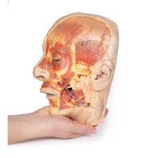
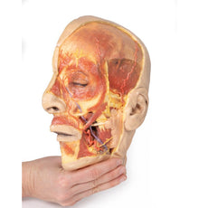
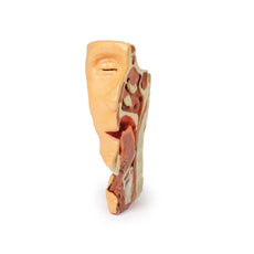
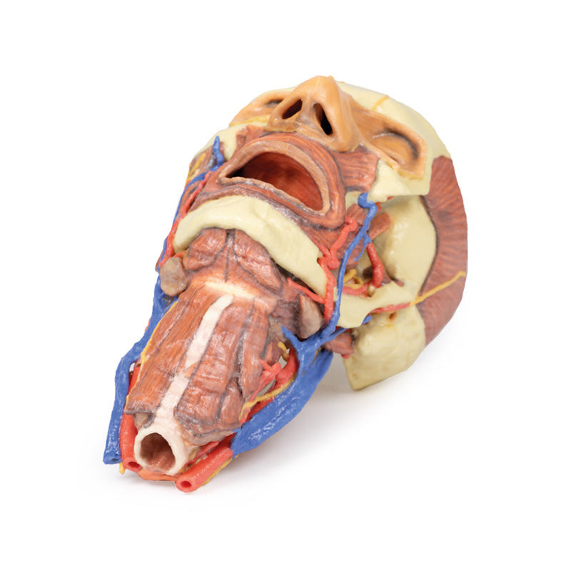
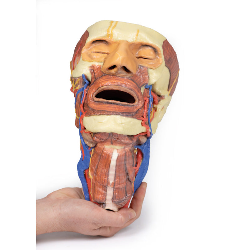
Viewed from the anterior aspect the face has been dissected to display some of the facial muscles around the mouth (buccinator [on the left], orbicularis oris and zygomaticus major). On the left side of the infratemporal fossa has been open to expose the medial and lateral pterygoids.
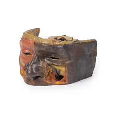
The lateral pterygoid is divided to show the mandibular division of the trigeminal nerve dividing into the lingual nerve and the inferior alveolar branch. Also on the left side the branches of the ophthalmic division of the trigeminal that supply the skin above the eyebrows and scalp (supraorbital [left only] and supratrochlear nerves [both sides]) are dissected. The submandibular gland is clearly visible below the mandible on both sides as are the facial arteries and veins as they course over the mandible.
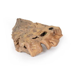
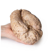
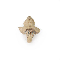
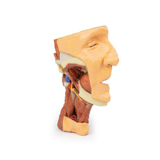
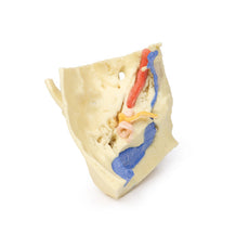
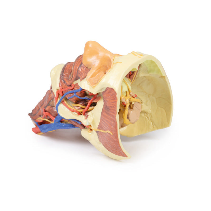
The neck: The musculoskeletal portion of the neck have been removed to display the pharynx posteriorly, the larynx anteriorly, and the neurovascular bundles laterally. The suprahyoid and infrahyoid muscles can be seen on the neck, as well as the cricothyroid muscle.
When looking up the length of the trachea from below, the vocal folds are visible. The hypoglossal nerve can be seen winding around the lateral surface of the external carotid artery and the external branch of superior laryngeal nerve is seen descending in the neck. The internal jugular vein, the common carotid artery and its bifurcation into external and internal carotid arteries are clearly seen on both left and right. The vagus nerve in the carotid sheath is also visible. The ansa cervicalis is visible emerging below the digastric muscle and descending on the surface of the internal jugular vein. The internal branch of the superior laryngeal nerve can be seen below the superior thyroid artery on the left. The superior thyroid artery branching from the external carotid artery is seen descending in the anterior neck. The internal branch of the superior laryngeal artery is visible on the left piercing the thyrohyoid membrane above the inferior constrictor where this muscle is attached to the hyoid bone.
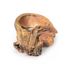
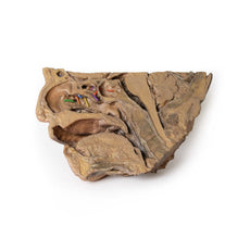
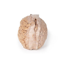
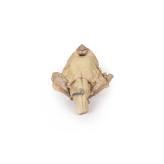
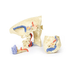
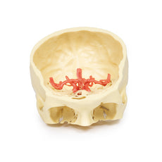
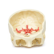
Posterior view of the pharynx: The superior, middle and inferior pharyngeal constrictors are indicated on the pharynx wall. The oesophagus can be identified emerging from the lower end of the pharynx. The posterior horn of the hyoid bone acts as a useful landmark. The carotid sheath seen from behind clearly shows the vagus nerve and its pharyngeal branches on the left. The recurrent laryngeal nerve is briefly visible on the left lying medial to the inferior thyroid artery. The occipital arteries are visible as they curve around the mastoid process. The vertebral arteries are seen either side of the brainstem as they enter the foramen magnum. The cerebellum has been removed to allow the fourth ventricle to be exposed. The cut surfaces of the cerebellar peduncles are clearly visible. A large portion of the posterior inferior cerebellar artery on the right is still visible as it winds around around the brainstem.
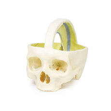
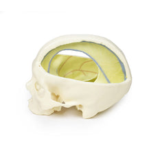
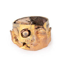
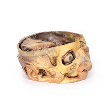
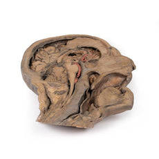
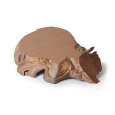
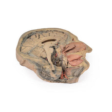
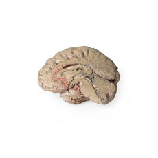
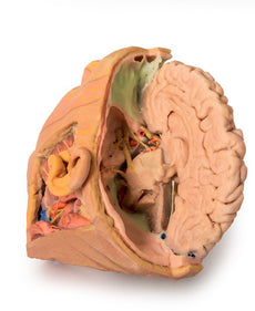
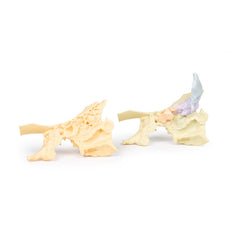
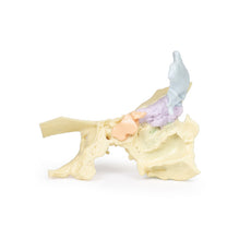
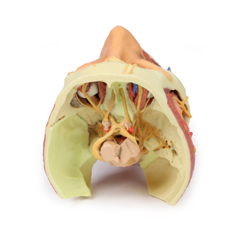
Cranial Cavity: The left and right orbits have been opened to reveal the orbital nerves and vessels along with the eyes and optic nerves. The optic chiasm, optic tracts and the lateral geniculate bodies are retained thus showing a large part of the visual pathways. The brainstem is cut at the level of the superior colliculi on the left and slightly lower on the right. The olfactory tracts and bulbs are also demonstrated. The origins of many of the cranial nerves from the brainstem are clearly visible.
Download Handling Guidelines for 3D Printed Models
GTSimulators by Global Technologies
Erler Zimmer Authorized Dealer


3D Printed Head and Visceral Column of the Neck
Item # MP1670
$2,723.00
$3,026.00
You save $303.00
Need an estimate?
Click Add To Quote

Features & Specifications
-
 by
by
A trusted GT partner -
FREE Shipping
U.S. Contiguous States Only -
 3D Printed Model
from a real specimen -
 Gov't pricing
Available upon request
 by
by 
Frequently Bought Together

3D Printed Head and Visceral Column of the Neck
This 3D print specimen preserves a series of features of the head and visceral column of the neck:The face: On the right side of the head the parotid gland has been removed to reveal the facial nerve and all its branches (temporal, zygomatic, buccal, marginal mandibular and cervical) and demonstrate the spatial relations of structures embedded in the gland from superficial to deep (facial nerve, retromandibular vein, external carotid artery). In the surrounding region the temporalis, masseter and posterior belly of digastric are exposed, as are and the facial artery, transverse facial artery and superficial temporal artery. The facial vein and transverse facial vein are clearly visible uniting to form the common facial vein which is joined by the retromandibular vein to form the external jugular vein.
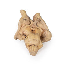
Viewed from the anterior aspect the face has been dissected to display some of the facial muscles around the mouth (buccinator [on the left], orbicularis oris and zygomaticus major). On the left side of the infratemporal fossa has been open to expose the medial and lateral pterygoids.
The lateral pterygoid is divided to show the mandibular division of the trigeminal nerve dividing into the lingual nerve and the inferior alveolar branch. Also on the left side the branches of the ophthalmic division of the trigeminal that supply the skin above the eyebrows and scalp (supraorbital [left only] and supratrochlear nerves [both sides]) are dissected. The submandibular gland is clearly visible below the mandible on both sides as are the facial arteries and veins as they course over the mandible.
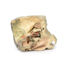
The neck: The musculoskeletal portion of the neck have been removed to display the pharynx posteriorly, the larynx anteriorly, and the neurovascular bundles laterally. The suprahyoid and infrahyoid muscles can be seen on the neck, as well as the cricothyroid muscle.
When looking up the length of the trachea from below, the vocal folds are visible. The hypoglossal nerve can be seen winding around the lateral surface of the external carotid artery and the external branch of superior laryngeal nerve is seen descending in the neck. The internal jugular vein, the common carotid artery and its bifurcation into external and internal carotid arteries are clearly seen on both left and right. The vagus nerve in the carotid sheath is also visible. The ansa cervicalis is visible emerging below the digastric muscle and descending on the surface of the internal jugular vein. The internal branch of the superior laryngeal nerve can be seen below the superior thyroid artery on the left. The superior thyroid artery branching from the external carotid artery is seen descending in the anterior neck. The internal branch of the superior laryngeal artery is visible on the left piercing the thyrohyoid membrane above the inferior constrictor where this muscle is attached to the hyoid bone.
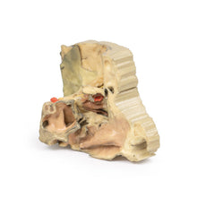
Posterior view of the pharynx: The superior, middle and inferior pharyngeal constrictors are indicated on the pharynx wall. The oesophagus can be identified emerging from the lower end of the pharynx. The posterior horn of the hyoid bone acts as a useful landmark. The carotid sheath seen from behind clearly shows the vagus nerve and its pharyngeal branches on the left. The recurrent laryngeal nerve is briefly visible on the left lying medial to the inferior thyroid artery. The occipital arteries are visible as they curve around the mastoid process. The vertebral arteries are seen either side of the brainstem as they enter the foramen magnum. The cerebellum has been removed to allow the fourth ventricle to be exposed. The cut surfaces of the cerebellar peduncles are clearly visible. A large portion of the posterior inferior cerebellar artery on the right is still visible as it winds around around the brainstem.
Cranial Cavity: The left and right orbits have been opened to reveal the orbital nerves and vessels along with the eyes and optic nerves. The optic chiasm, optic tracts and the lateral geniculate bodies are retained thus showing a large part of the visual pathways. The brainstem is cut at the level of the superior colliculi on the left and slightly lower on the right. The olfactory tracts and bulbs are also demonstrated. The origins of many of the cranial nerves from the brainstem are clearly visible.
Download Handling Guidelines for 3D Printed Models
GTSimulators by Global Technologies
Erler Zimmer Authorized Dealer
These items normal warranty are two years, however the warranty doesn’t cover “wear and tear”. The manufacturer does have 100% quality control on these models.
The models are very detailed and delicate. With normal production machines you cannot realize such details like shown in these models.
The printer used is a color-plastic printer. This is the most suitable printer for these models.
The plastic material is already the best and most suitable material for these prints. (The other option would be a kind of gypsum, but this is way more fragile. You even cannot get them out of the printer without breaking them).The huge advantage of the prints is that they are very realistic as the data is coming from real human specimen. Nothing is shaped or stylized.
The users have to handle these prints with utmost care. They are not made for touching or bending any thin nerves, arteries, vessels etc. The 3D printed models should sit on a table and just rotated at the table.
Related Products


$2,784.00
$3,094.00
Free shipping
3D Printed Sagittal Section of Head with Infratemporal Fossa Dissection
Item # MP1104

$1,189.00
$1,322.00
Free shipping
3D Printed Parotid Gland and Facial Nerve Dissection
Item # MP1112

$2,914.00
$3,238.00
Free shipping
3D Printed Sagittal Section of Head and Neck with Infratemporal Fossa and Carotid Sheath Dissection
Item # MP1111


$2,329.00
$2,588.00
Free shipping
3D Printed Superficial Facial Nerves & Parotid Gland
Item # MP1109


$2,799.00
$3,110.00
Free shipping
3D Printed Parasagittal Section of the Head and Neck
Item # MP1107


$2,185.00
$2,428.00
Free shipping
3D Printed Median Section Through Head Sagittal Section of Head with Deep Dissection
Item # MP1105


$473.00
$526.00
3D Printed Brain Stem, Isolated Anatomy From Midbrain to Medulla Oblongata
Item # MP1101


$8,528.00
$9,476.00
Free shipping
3D Printed Head, Neck, Shoulder and Thorax Replica with Angiosomes
Item # MP1250
by — Item # MP1670
3D Printed Head and Visceral Column of the Neck
$2,723.00
$3,026.00
Add to Cart
Add to Quote
