Your shopping cart is empty.
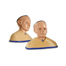
Enhanced Digital Eye and Ear Examination Trainer Set, Light


Normal Ear
Outer Ear
Outer / Middle Ear
Middle Ear
Skull Base

 Ear Product's Manual
Eye Product's Manual


0.0 lb


Enhanced Digital Eye and Ear Examination Trainer Set, Light
Item # AR505
$5,382.00
$5,666.00
You save $284.00
In stock
Need an estimate?
Click Add To Quote

Features & Specifications
-
 by
by
A trusted GT partner -
FREE Shipping
U.S. Contiguous States Only -
2-Year Warranty
Provided by manufacturer -
 Gov't pricing
Available upon request
 by
by Frequently Bought Together

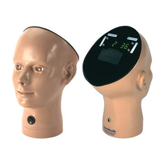
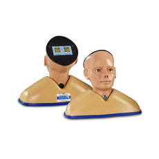
Enhanced Digital Eye and Ear Examination Trainer Set, Light
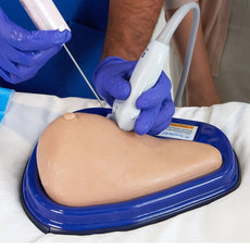
The Enhanced Eye and Ear Examination Trainer Set brings together two cutting-edge simulation tools, the Enhanced
Digital
Ear Examination Trainer and the Enhanced Digital Eye Examination Trainer, to deliver a comprehensive, and inclusive
learning experience in clinical examination skills.
Developed in collaboration with Professor Vinod Patel, Professor, Diabetes and Clinical Skills and Professor Tony
Wright, Emeritus Professor of Otolaryngology, this dual trainer set redefines realism in otoscopy and ophthalmoscopy
training.
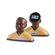
The Enhanced Ear model features a comprehensive, newly classified library of 59 ear conditions, displayed in
lifelike
detail on a high-resolution circular LCD screen. Representations of the tympanic membrane are the same in both light
and
dark skin toned models.
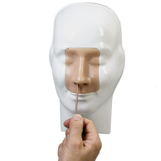
Using the Enhanced Eye model trainees can explore 36 eye conditions displayed in vivid clarity on a high-resolution
circular LCD screens.
In darker-skinned patients, deeper pigmentation in the fundus reduces vessel visibility, a nuance replicated only in
the
AR503/50 Dark model. Offering two product versions enables trainees to gain an awareness of these visible variances.
Both products feature a responsive touchscreen interface with intuitive control, allowing users to browse, search,
randomise, or display conditions in customisable sets. Dedicated Exam and Sleep modes allow for flexible training or
assessment scenarios.
Together, the Set offers a seamless, advanced platform for mastering ear and eye examination techniques, combining
anatomical realism, digital precision, and inclusive representation to elevate the standard of clinical education
worldwide.
- Condition Library: browse names and image previews for all conditions
- Search Function: instantly locate a condition for teaching or self-directed study
- Random Mode: display conditions in a varied sequence to keep learners interested
- Custom Sets: select up to 10 specific conditions in any order, for focused teaching or assessments
- Exam Mode: hide condition names and numbers for authentic test conditions. Sleep Mode is automatically disabled for uninterrupted practice
- Compatible with battery or USB-C mains power
- Intelligent sleep mode for power saving (automatically cancelled in Exam Mode)
Features
Digital Ear Conditions
Ear conditions and diseases presented digitally for the right patient ear:- 1 - Normal I
- 2 - Normal II
- 3 - Ear Wax (Cerumen)
- 4 - Swimmer's Osteoma
- 5 - Fungal Ear I
- 6 - Fungal Ear II
- 7 - Foreign Body I
- 8 - Foreign Body II
- 9 - Acute Haemorrhagic Bullous Otitis Externa
- 10 - Viral Papilloma
- 11 - External Canal Stenosis
- 12 - Furunculosis
- 13 - Skull Base Fracture
- 14 - Pseudomonas Otitis Externa
- 15 - Keratosis Obturans
- 16 - Barotrauma I
- 17 - Barotrauma II
- 18 - Acute Viral Ear
- 19 - Acute Secretory Otitis Media I
- 20 - Resolving Secretory Otitis Media
- 21 - Acute Secretory Otitis Media II
- 22 - Acute Otitis Media III
- 23 - Perforation Following an Acute Suppurative Otitis Media (ASOM)
- 24 - Childhood Glue Ear
- 25 - Glue Ear in a Child with Dermoid Cyst in the Eardrum
- 26 - Adult Glue Ear
- 27 - A Standard Ventilation Tube in the Membrane
- 28 - Infected Mini Grommet with Otitis Externa Secondary to a Mucus Discharge
- 29 - Permanent Ventilation Tube in Place
- 30 - Large Perforation of the Tympanic Membrane
- 31 - A Posterior Perforation of the Tympanic Membrane
- 32 - Two Small Traumatic Perforations Following a Blow to the Ear
- 33 - Subtotal Perforation of the Tympanic Membrane
- 34 - Chronic Suppurative Otitis Media-Mucosal
- 35 - Perforation with Tympanosclerosis
- 36 - Grommet Scar Healed
- 37 - Foreign Body in Middle Ear
- 38 - Tympanosclerosis of Tympanic Membrane
- 39 - Posterior Retraction
- 40 - Retraction onto Long Process of the Incus
- 41 - Retraction with Loss of Long Process of the Incus and Keratin Trail
- 42 - Retraction with Loss of Long Process of the Incus
- 43 - Posterior Retraction Pocket onto Jugular Bulb and with Middle Ear Fluid
- 44 - Retraction with Early Keratin Build Up
- 45 - Childhood Attic Retraction
- 46 - Deep Attic Retraction
- 47 - Attic Retraction Accumulation Keratin Underlying Cholesteatoma
- 48 - Extensive Accumulation with Cholesteatoma in Middle Ear
- 49 - Wet Cholesteatoma
- 50 - Aural Polyp
- 51 - Clean Dry Reconstructed Mastoid Cavity
- 52 - Old Style Mastoid Cavity with Residual Cholesteatoma
- 53 - Mastoid Cavity with Fistula into Lateral Semicircular Canal
- 54 - Congenital Cholesteatoma
- 55 - Large Congenital Cholesteatoma
- 56 - Ear Canal Cholesteatoma I
- 57 - Ear Canal Cholesteatoma II
- 58 - Glomus Tympanicum Tumours
- 59 - Glomus Jugulare Tumour
Digital Eye Conditions
Eye conditions and diseases presented digitally for the light skin toned patients AR503 and dark skin toned patients AR503/50
-
Diabetic Retiopathy:
- 1 - Background Diabetic Retinopathy (R1) with Maculopathy (M0)
- 2 - Pre-Proliferative Diabetic Retinopathy (R2)
- 3 - Proliferative Diabetic Retinopathy (R3)
- 4 - Proliferative Diabetic Retinopathy - New Vessels on Optic Disc (R3)
- 5 - Diabetic Maculopathy (M2)
- 6 - Diabetic Maculopathy (M2) with Laser Photocoagulation (P1)
- 7 - Laser Photocoagulation (P1) with Proliferative Diabetic Retinopathy (R3) at the Optic Disc
- 8 - Ungradable - Proliferative Retinopathy Likely - (R3)
-
Important/Common Retinal Conditions:
- 9 - Normal Fundus (Optic Disc and Retina)
- 10 - Glaucoma
- 11 - Papilloedema
- 12 - Optic Atrophy with Macular Scarring (and Glaucoma)
- 13 - Early Dry Age-Related Macular Degeneration
- 14 - Hypertensive Retinopathy: Grade 2
- 15 - Central Retinal Vein Occlusion (CRVO)
- 16 - Central Retinal Artery Occlusion (CRAO)
- 17 - Drusen
- 18 - Retinitis Pigmentosa
- 19 - Myelinated Nerve Fibres
- 20 - High Myopia
- 21 - Branch Retinal Vein Occlusion (BRVO)
- 22 - Pre-Retinal Haemorrhage
-
Important/Less Common Retinal Conditions:
- 23 - Multiple Retinal Haemorrhages
- 24 - Retinal Detachment
- 25 - Angioid Streaks
- 26 - Benign Disc Neavus
- 27 - Malignant Melanoma
- 28 - Macular Haemorrhage
- 29 - Choroidal Naevus
- 30 - Macular Scar (Toxoplasmosis)
- 31 - Cytomegalovirus Retinitis
- 32 - Lipaemia Retinalis with Proliferative Diabetic Retinopathy
- 33 - Medusa Head Optic Disc (This is Normal Variant)
- 34 - Myopic Crescent - Normal Choroidal Vessels
- 35 - A Resolving Pre-Retinal Haemorrhage
- 36 - Macular Burn with Peripheral Burn with Scarring
Includes
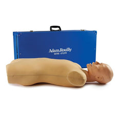
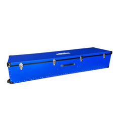
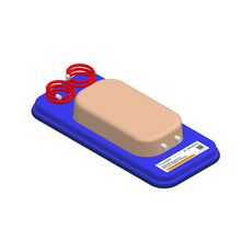
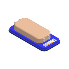
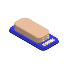
- Rigid Carrying Case (x2)
- Shoulder Base (x2)
- USB-C Mains Adaptor with Worldwide Plug Fixings (x2)
- Instruction Manual (x2)
Ear Product's Manual
Eye Product's Manual
GTSimulators by Global Technologies
Adam-Rouilly Authorized Dealer
Enhanced Digital Eye and Ear Examination Trainer Set, Light
-
Enhanced Digital Ear Examination Dimensions and Weight:
- Dimensions: 17 x 11 x 19 in
- Weight: 12 lbs
-
Enhanced Digital Eye Examination Dimensions and Weight:
- Dimensions: 17 x 11 x 19 in
- Weight: 10 lbs
GTSimulators by Global Technologies
Adam-Rouilly Authorized Dealer
Related Products


$5,449.00
Was $5,736.00
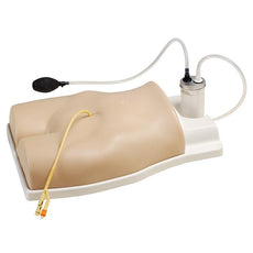
CORMAN - Adult Nasogastric and Nasojejunal Feeding Trainer, Light Skin
Item # AR90


$1,285.00
Was $1,353.00
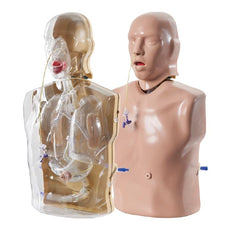
Injection, Venipuncture, Cannulation, and Infusion Training Arm
Item # AR251


$1,952.00
Was $2,055.00
Carrying Case For Full Body X-Ray and Radiographic Positioning Manikin (AR10A)
Item # ARR03001


$5,449.00
Was $5,736.00
CORMAN - Adult Nasogastric and Nasojejunal Feeding Trainer, Dark Skin
Item # AR90/50


by — Item # AR505
Enhanced Digital Eye and Ear Examination Trainer Set, Light
$5,382.00
$5,666.00
Add to Cart
Add to Quote