Your shopping cart is empty.
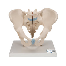
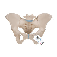
Male pelvis with ligaments, pelvic floor and organs, 7-parts

 Independence Day Savings

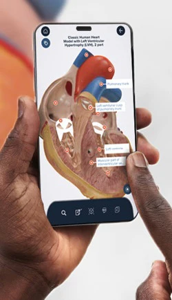
Every original 3B Scientific® Anatomy Model gives you direct access to its digital twin.
Enjoy using the exclusive virtual anatomy content with the following features:
1. Freely rotate your digital model and zoom in and out
2. Display hotspots and their anatomical structures
3. Augmented Reality (AR) feature starts your virtual anatomy model
4. Anatomy Quiz function to test and improve your anatomical knowledge with instant results and final score evaluation
5. Drawing function that allows image customization with save and share function
6. Useful Notes function to help you with your personal learning
7. Possibility to learn both male and female anatomy
8. Easy access to 3D content both online and offline
9. Available in 11 languages
To get started, simply scan the QR-code located on your 3B Scientific® Anatomical Model, download the new 3B Smart Anatomy app and step into the virtual world of Human Anatomy.
Learn more 3B Smart Anatomy.


0.0 lb


This product includes FREE access to the 3B Smart Anatomy course — Learn more
Independence Day Savings
Discount has been applied automatically for this item.
Male pelvis with ligaments, pelvic floor and organs, 7-parts
Item # H21/3
$844.00
$998.00
You save $154.00
Subject to Availability
Need an estimate?
Click Add To Quote

Features & Specifications
-
 by
by
A trusted GT partner -
FREE Shipping
U.S. Contiguous States Only -
3-Year Warranty
Provided by manufacturer -
 3B Smart Anatomy
Free access to the course -
 Gov't pricing
Available upon request
 by
by 
Frequently Bought Together

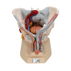
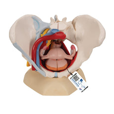
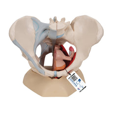
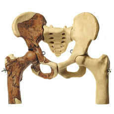
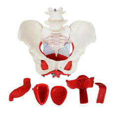
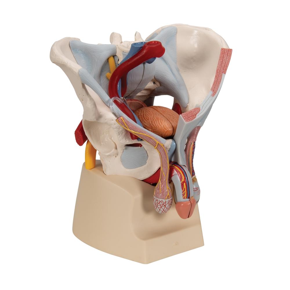
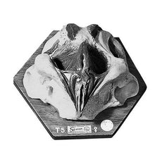
Male pelvis with ligaments, vessels, nerves, pelvic floor and organs, 7-parts - Includes 3B Smart Anatomy
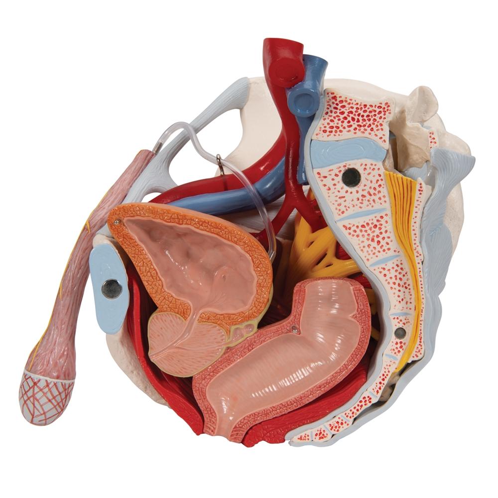
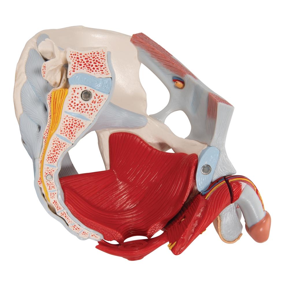
This 7 part model of the male pelvis shows in accurate detail how the bones, ligaments, vessels and nerves as well
as
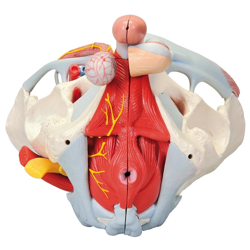
the pelvic floor muscles and the external sex organs are connected to each other. It shows the whole pelvis, through
which a median section has been placed. The right side of the external anal sphincter, the M. ischiocavernosus, the
M.
transversus perinei profundus and superficialis and the M. bulbospongiosus can be removed together.
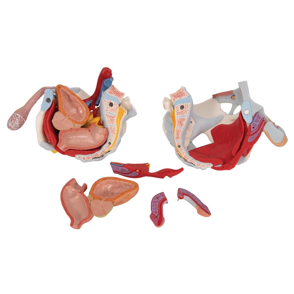
The rectum, bladder, prostate and penis can also be removed, and split into two halves at median level - partially
connected. The bone structures are connected with magnets and can therefore be easily taken apart. The skin and the
fascia of the penis have been partly removed so that the vessels and nerves are visible. Part of the skin in the
area of
the scrotum and the spermatic cord has removed too. The testicles and epididymis are also visible. On the left, the
spermatic cord has been opened up in layers, and on the right the M. cremaster and fascia spermatica interna have
been
exposed.
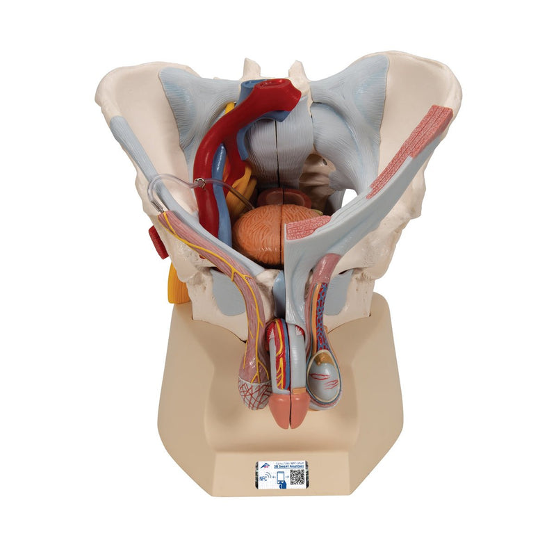
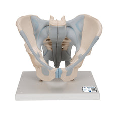
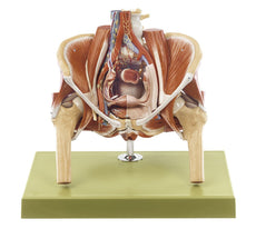
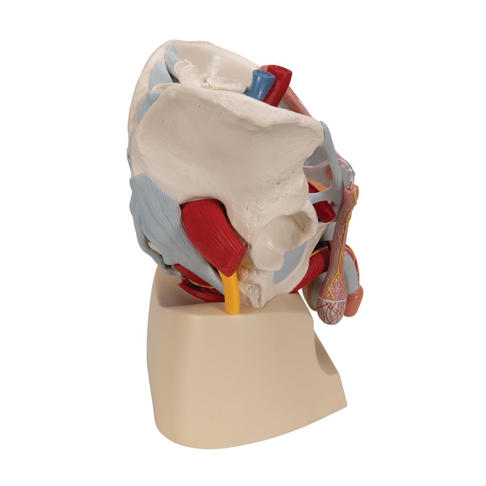
The right half of the pelvis shows sections of the common, external and internal iliac arteries and the common and
external iliac veins, demonstrating their positions in relation to each other. The Plexus sacralis, the N.
ischiadicus,
the N. pudendus, the N. dorsalis penis, the Nn. scrotales anteriores, the Nn. perineales and the Nn. annales
inferiores
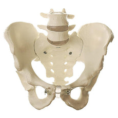
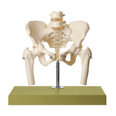
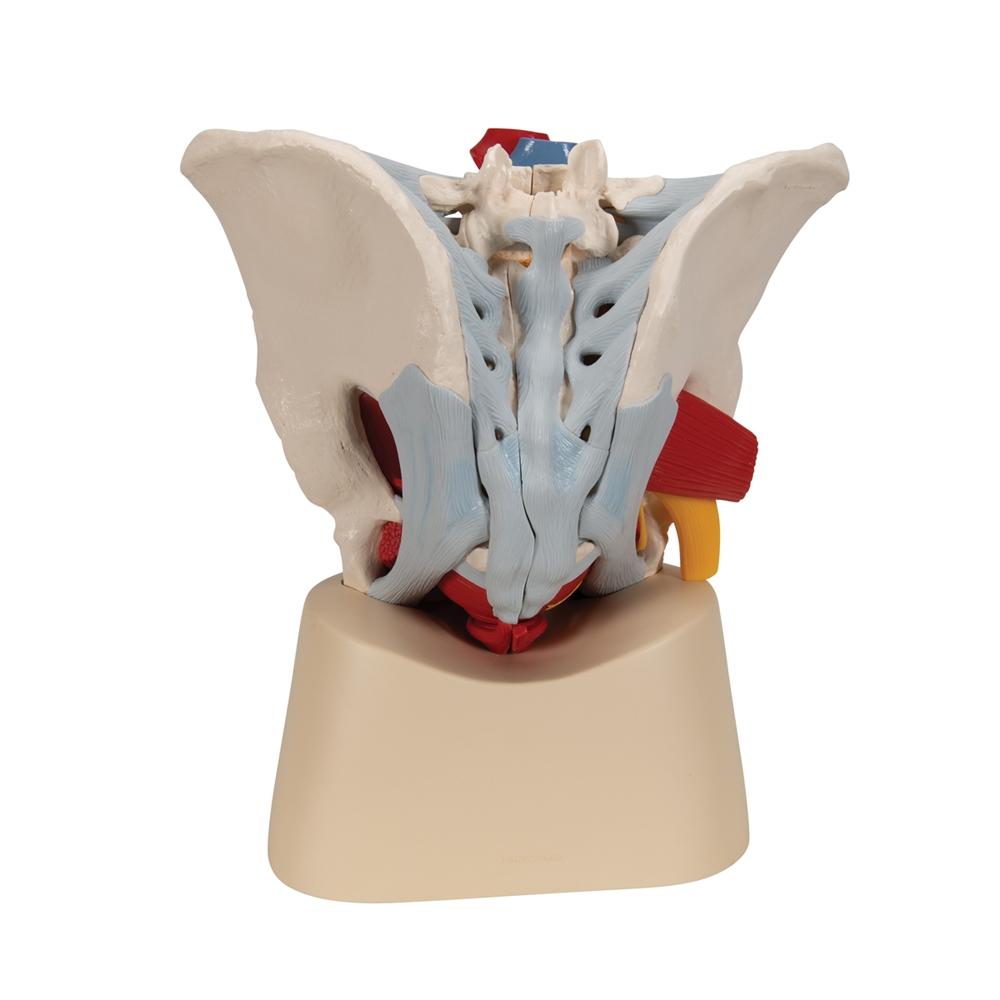
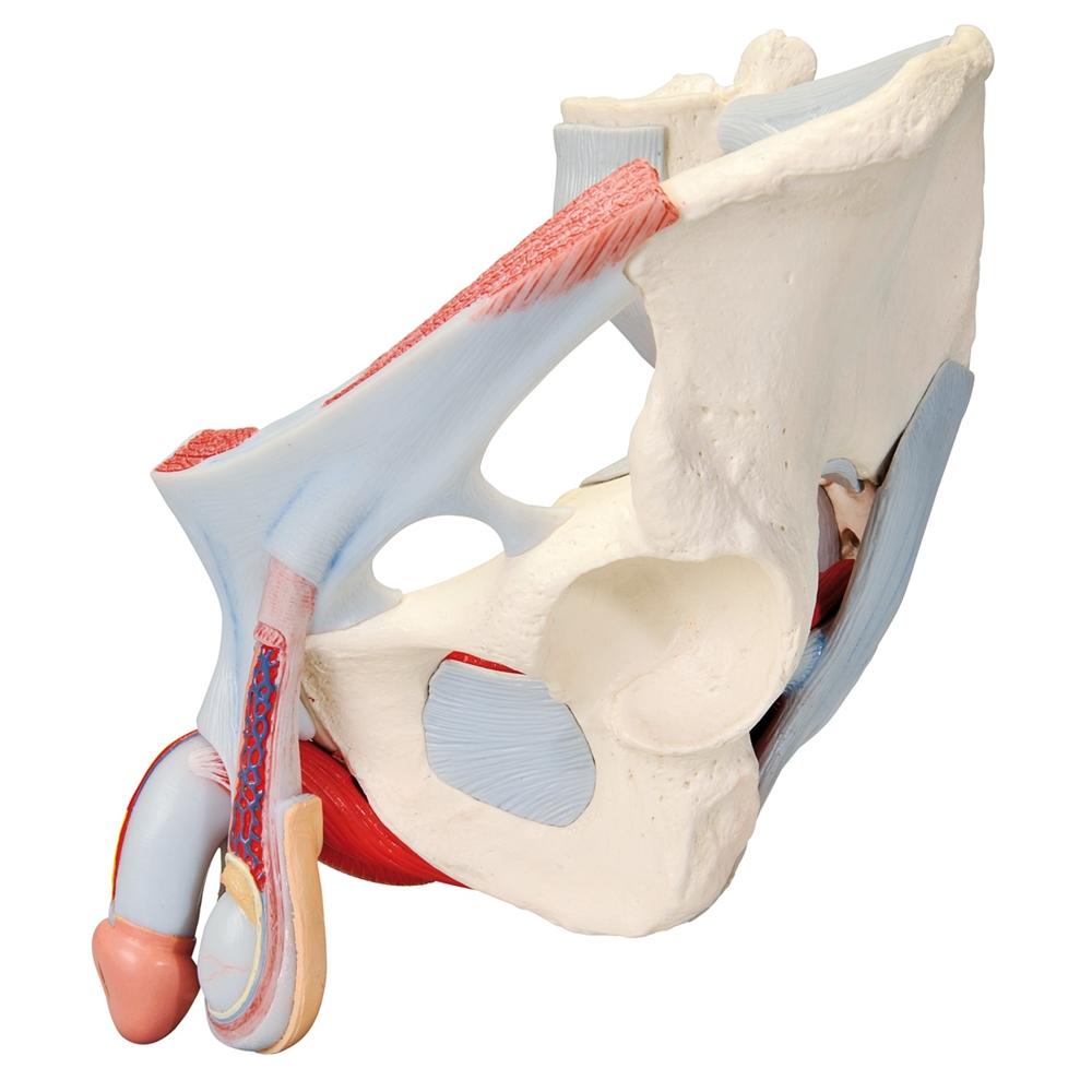
as well as the Ductus deferens are shown. The model shows the following bones and ligaments: both hip bones, pubic
symphisis, sacrum and coccyx as well as the fifth lumbar vertebra with intervertebral disc. A median section has
been
placed through the fifth lumbar vertebra, the sacrum and the coccyx, so that the pelvis can be split into two
halves.
This means that part of the cauda equina is also visible in the vertebral canal. The left half of the fifth lumbar
vertebra can be removed.
-
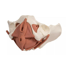
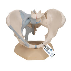
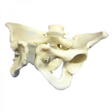
The following ligament structures of the pelvis are shown:
- Lig. inguinale
- Lig. sacrotuberale
- Lig. sacrospinale
- Lig. sacroiliaca anteriora
- Lig. iliolumbale
- Lig. longitudinale anterius
- Lig. Supraspinale, Lig. sacroiliacum interosseum
- Lig. sacroiliacum posterius
- Lig. sacrococcygeum laterale
- Lig. sacrococcygeum posterius superficiale et profundum Membrana obturatoria
- Lig. lacunare
GTSimulators by Global Technologies
3B Scientific Authorized Dealer.

Every original 3B Scientific® Anatomy Model gives you direct access to its digital twin.
You can use it on your smartphone, tablet or desktop device.
Enjoy using the exclusive virtual anatomy content with the following features:
1. Freely rotate your digital model and zoom in and out
2. Display hotspots and their anatomical structures
3. Augmented Reality (AR) feature starts your virtual anatomy model
4. Anatomy Quiz function to test and improve your anatomical knowledge with instant results and final score evaluation
5. Drawing function that allows image customization with save and share function
6. Useful Notes function to help you with your personal learning
7. Possibility to learn both male and female anatomy
8. Easy access to 3D content both online and offline
9. Available in 11 languages
To get started, simply scan the QR-code located on your 3B Scientific® Anatomical Model, download the new 3B Smart Anatomy app and step into the virtual world of Human Anatomy.
Learn more 3B Smart Anatomy.
Male pelvis with ligaments, vessels, nerves, pelvic floor and organs, 7-parts - Includes 3B Smart Anatomy
Product Dimensions and Weight:- Size: 8.3 x 11.0 x 12.2 in
- Weight: 6.88 lb
GTSimulators by Global Technologies
3B Scientific Authorized Dealer.
Related Products


$814.00
Was $963.00
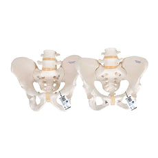
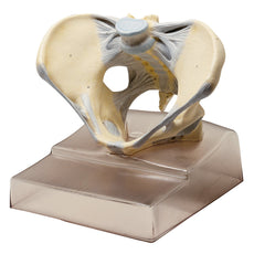
Female Pelvis Model with Ligaments, Pelvic Floor and Organs, 6 part
Item # H20/4


by — Item # H21/3
Male pelvis with ligaments, pelvic floor and organs, 7-parts
$844.00
$998.00
Add to Cart
Add to Quote