

Your shopping cart is empty.
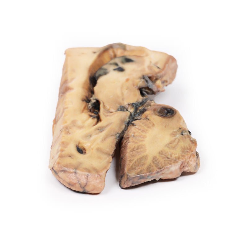
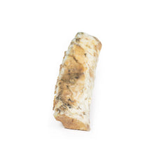
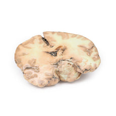
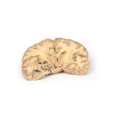
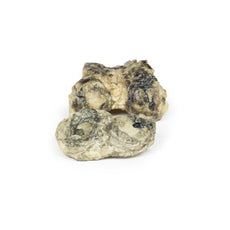
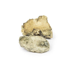
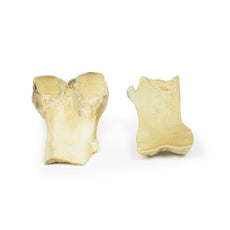
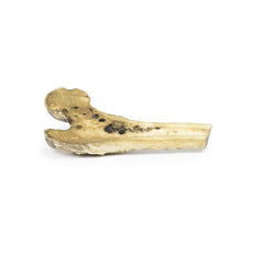
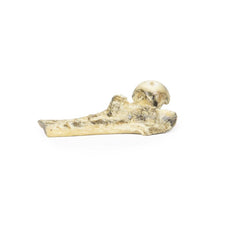
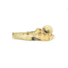
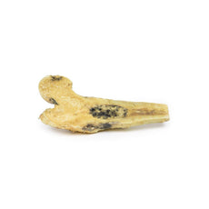
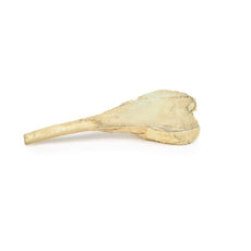
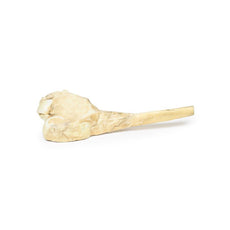
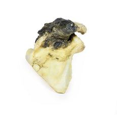
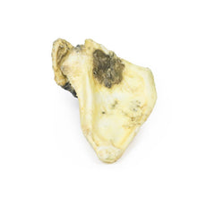
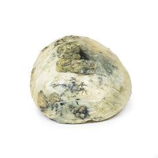
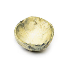
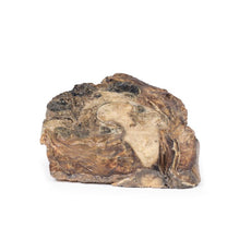
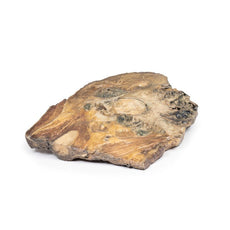
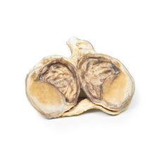
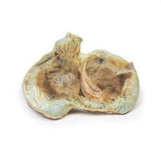
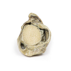
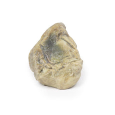
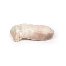
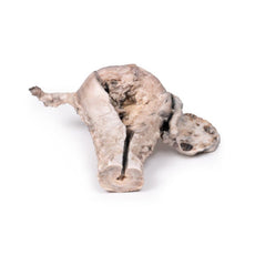
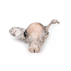
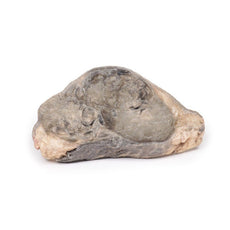
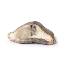
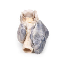
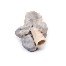
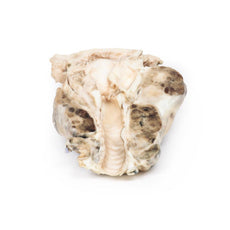
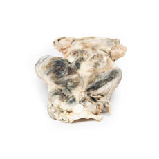
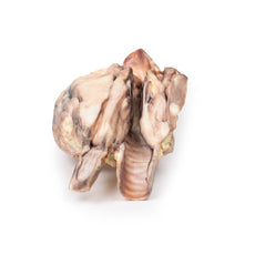
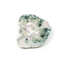
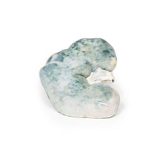
3D Printed Berry Aneurism of Basilar Artery

 Handling Guidelines for 3D Printed Models
GTSimulators by Global Technologies
Erler Zimmer Authorized Dealer
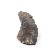
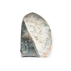
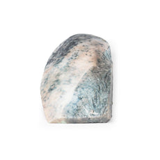


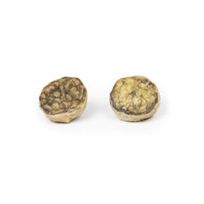
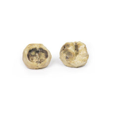

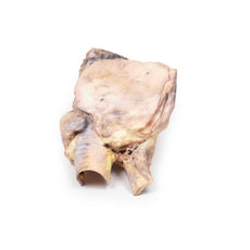


3D Printed Berry Aneurism of Basilar Artery
Item # MP2001
$385.00
$429.00
You save $44.00
Need an estimate?
Click Add To Quote

Features & Specifications
-
 by
by
A trusted GT partner -
 3D Printed Model
from a real specimen -
 Gov't pricing
Available upon request
 by
by 
Frequently Bought Together

3D Printed Berry Aneurism of Basilar Artery
Clinical History
A 37-year-old patient presented to hospital after falling and striking his head, with
subsequent symptoms of headache, vomiting and disorientation. CT scan showed dilatation of the lateral
ventricles associated with a large mass projecting into the third ventricle posteriorly. One week later a shunt
was performed for hydrocephalus. An angiogram revealed a partially thrombosed aneurysm, measuring 1 x 1 cm,
arising from the basilar artery. At 3-months post-op the shunt was revised due to obstruction, with repeat
cerebral angiogram revealing interval enlargement of the aneurysm. Attempted ligation of the aneurysm was
unsuccessful. The patient remained unconscious despite several attempts to revise the shunt and he died.
Pathology
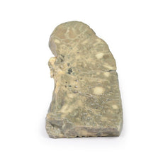
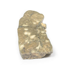
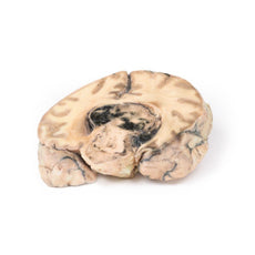
This brain has been sliced in the mid-sagittal plane. It comprises a whole hemi-section of the brain
about 1cm thick. On the medial surface a large darkly-coloured ovoid berry aneurysm measuring 5 x 2 cm in
diameter, arising from the basilar artery is clearly visible. It has eroded up into the midbrain, compressing
the third ventricle from below, and inferiorly into the substance of the pons. The wall of the aneurysm appears
intact although blood clot is seen in the third ventricle and appears to be leaking through the lateral wall of
that ventricle. The aneurysm is filled with a laminated thrombus. A small area of mucoid degeneration measuring
0.4 cm in diameter is seen posterior to the aneurysm within the pons.
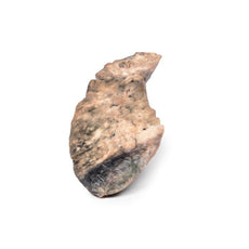
Examination of the lateral aspect of
the sagittal section shows dilatation of the lateral ventricle, blood staining of the ventricular wall and
patchy haemorrhagic infarction of the caudate nucleus. There was some discolouration of the meninges overlying
the tip of the left temporal lobe and the cerebellum (not included in 3D print), consistent with sub-arachnoid
haemorrhage.
Further Information
Prevalence of aneurysms is approximately 3.2% in the population, while rupture is much
less common, occurring only 7.9 per 100,000 person-years. A minority of intracranial aneurysms arise from the
posterior circulation, and are mostly situated at junctional points about the basilar, vertebral and cerebellar
arteries. Symptoms are either secondary to subarachnoid haemorrhage or a mass effect with associated compression
of the adjacent brain parenchyma and cranial nerves. Rupture causes complications due to bleeding and raised
intracranial pressure. Hydrocephalus, re-bleed and vasospasm may also occur. Management is via surgical means;
in recent years, novel therapies include endovascular intervention with coils and subsequent monitoring.
Handling Guidelines for 3D Printed Models
GTSimulators by Global Technologies
Erler Zimmer Authorized Dealer
These items normal warranty are two years, however the warranty doesn’t cover “wear and tear”. The manufacturer does have 100% quality control on these models.
The models are very detailed and delicate. With normal production machines you cannot realize such details like shown in these models.
The printer used is a color-plastic printer. This is the most suitable printer for these models.
The plastic material is already the best and most suitable material for these prints. (The other option would be a kind of gypsum, but this is way more fragile. You even cannot get them out of the printer without breaking them).The huge advantage of the prints is that they are very realistic as the data is coming from real human specimen. Nothing is shaped or stylized.
The users have to handle these prints with utmost care. They are not made for touching or bending any thin nerves, arteries, vessels etc. The 3D printed models should sit on a table and just rotated at the table.
Related Products


$743.00
$827.00
Free shipping
3D Printed Papillary Transitional Cell Carcinoma of the Renal Pelvis
Item # MP2099


by — Item # MP2001
3D Printed Berry Aneurism of Basilar Artery
$385.00
$429.00
Add to Cart
Add to Quote
