

Your shopping cart is empty.
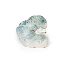
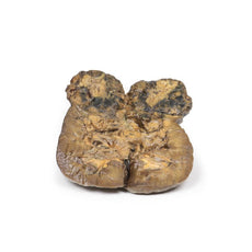
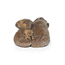
3D Printed Lobar Pneumonia

 Handling Guidelines for 3D Printed Models
GTSimulators by Global Technologies
Erler Zimmer Authorized Dealer
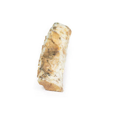
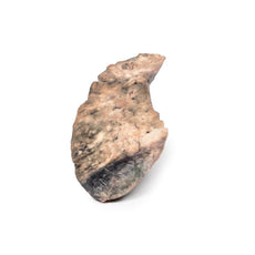
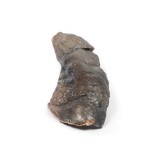
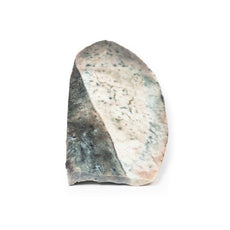
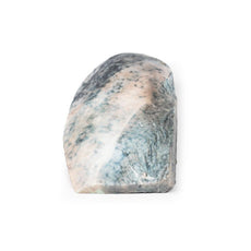


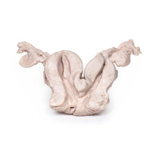
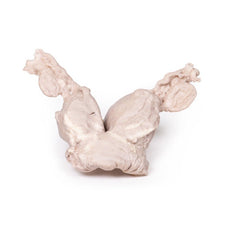

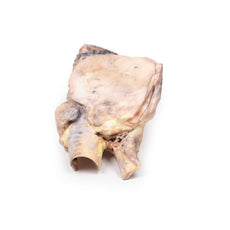


3D Printed Lobar Pneumonia
Item # MP2057
$939.00
$1,044.00
You save $105.00
Need an estimate?
Click Add To Quote

Features & Specifications
-
 by
by
A trusted GT partner -
FREE Shipping
U.S. Contiguous States Only -
 3D Printed Model
from a real specimen -
 Gov't pricing
Available upon request
 by
by 
Frequently Bought Together

3D Printed Lobar Pneumonia
Clinical History
There is no clinical history for this specimen.
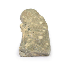
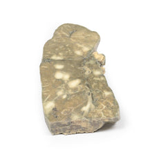
Pathology
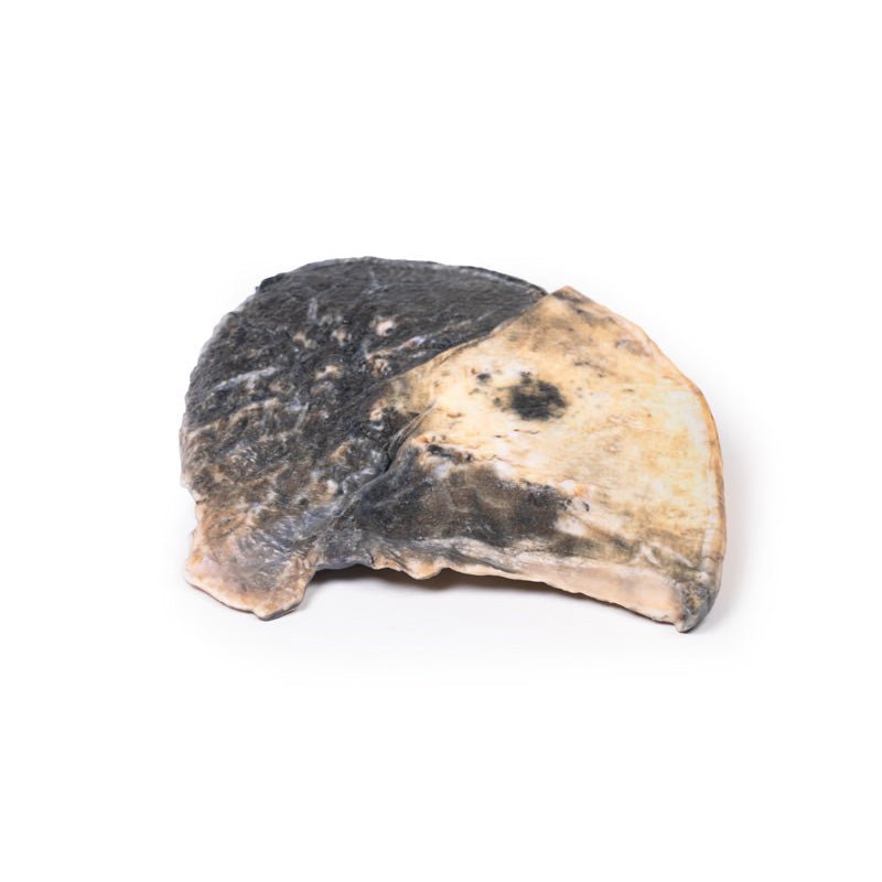
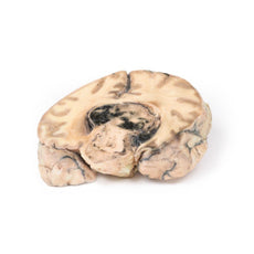
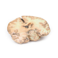
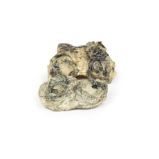
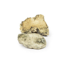
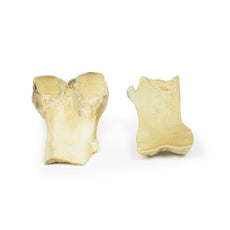
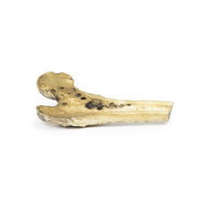
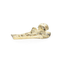
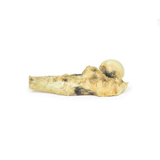
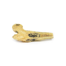
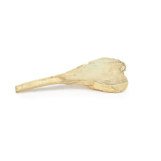
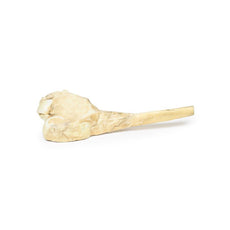
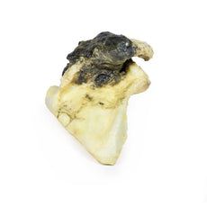
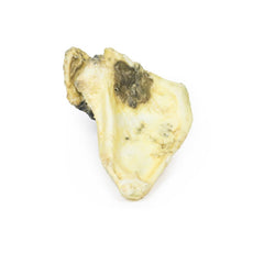
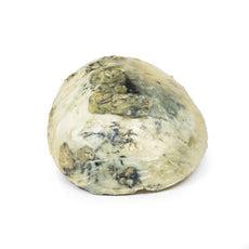
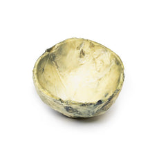
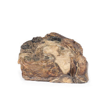
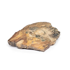
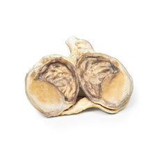
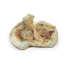
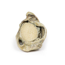
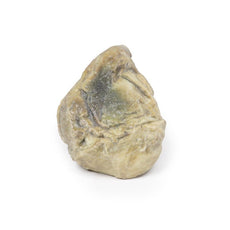
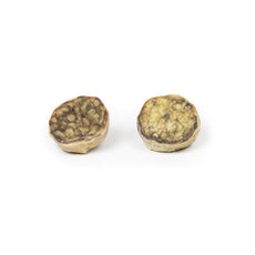
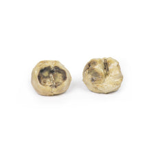
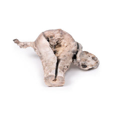
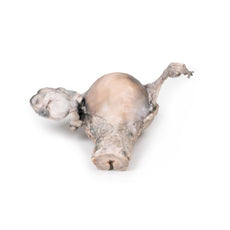
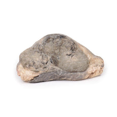
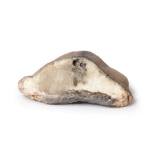
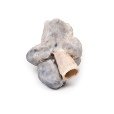
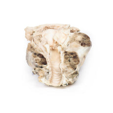
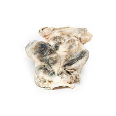
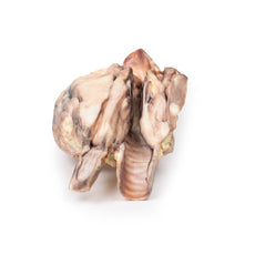
The specimen is a parasagittal section of the right lung and the boundaries between
the three lobes are visible. The entire upper and middle lobes are congested and hyperaemic* causing the darker
appearance. There are smaller foci in the left lung.
Further Information
Lobar pneumonia is a form of pneumonia characterized by inflammatory exudate
within the intra-alveolar space resulting in consolidation that affects a large and continuous area of the lobe of a
lung. It is one of the two anatomic classifications of pneumonia (the other being bronchopneumonia). The affected
lobe in this case shows classical red ‘hepatization’ or consolidation of the lung parenchyma, which is due to
vascular congestion with extravasation of red cells into alveolar spaces, along with increased numbers of
neutrophils and fibrin. The filling of the airspaces by the exudate leads to a gross appearance of solidification,
or consolidation, of the alveolar parenchyma. This reddish appearance has been likened to that of cut surface of the
liver, hence the term “hepatization”.
The most common organisms that cause lobar pneumonia are Streptococcus pneumoniae, also called pneumococcus, Haemophilus influenzae and Moraxella catarrhalis. Mycobacterium tuberculosis, the tubercle bacillus, may also cause lobar pneumonia if pulmonary tuberculosis is not treated promptly. Other organisms that lead to lobar pneumonia are Legionella pneumophila and Klebsiella pneumoniae.
Like other types of pneumonia, lobar pneumonia can present as a community-acquired infection, in immune suppressed
patients or as nosocomial infection. However, most causative organisms are of the community-acquired type.
On a
posteroanterior and lateral chest radiograph, an entire lobe will be radiopaque with no evidence of air within it,
indicative of lobar pneumonia.
*Hyperaemia = active engorgement of vascular beds with a normal or decreased
outflow of blood.
Handling Guidelines for 3D Printed Models
GTSimulators by Global Technologies
Erler Zimmer Authorized Dealer
These items normal warranty are two years, however the warranty doesn’t cover “wear and tear”. The manufacturer does have 100% quality control on these models.
The models are very detailed and delicate. With normal production machines you cannot realize such details like shown in these models.
The printer used is a color-plastic printer. This is the most suitable printer for these models.
The plastic material is already the best and most suitable material for these prints. (The other option would be a kind of gypsum, but this is way more fragile. You even cannot get them out of the printer without breaking them).The huge advantage of the prints is that they are very realistic as the data is coming from real human specimen. Nothing is shaped or stylized.
The users have to handle these prints with utmost care. They are not made for touching or bending any thin nerves, arteries, vessels etc. The 3D printed models should sit on a table and just rotated at the table.
Related Products


$743.00
$827.00
Free shipping
3D Printed Papillary Transitional Cell Carcinoma of the Renal Pelvis
Item # MP2099


by — Item # MP2057
3D Printed Lobar Pneumonia
$939.00
$1,044.00
Add to Cart
Add to Quote
