

Your shopping cart is empty.
3D Printed Meningioma

 Handling Guidelines for 3D Printed Models
GTSimulators by Global Technologies
Erler Zimmer Authorized Dealer
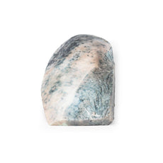


3D Printed Meningioma
Item # MP2004
$2,893.00
$3,215.00
You save $322.00
Need an estimate?
Click Add To Quote

Features & Specifications
-
 by
by
A trusted GT partner -
FREE Shipping
U.S. Contiguous States Only -
 3D Printed Model
from a real specimen -
 Gov't pricing
Available upon request
 by
by 
Frequently Bought Together

3D Printed Meningioma
Clinical History
A 68-year-old female presented with recent onset of seizures and was diagnosed with epilepsy.
Collateral history revealed a gradual change in the patient’s personality. She subsequently died several months
later from a myocardial infarction.
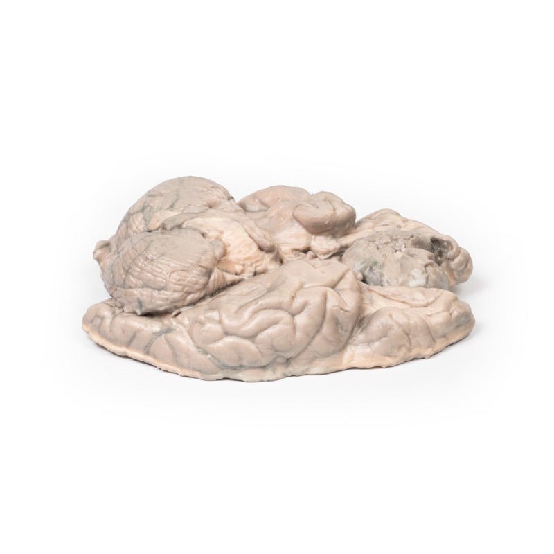
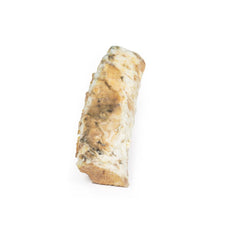
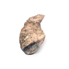
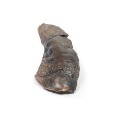
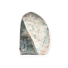
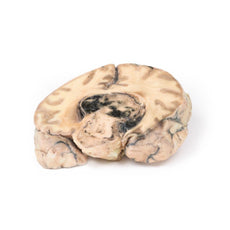
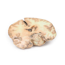
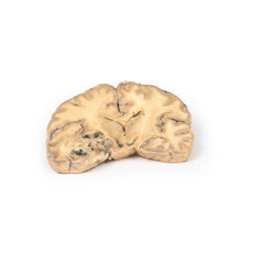
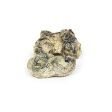
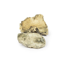
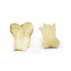
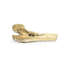
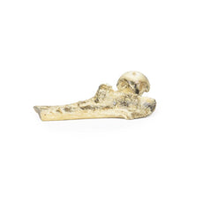
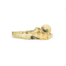
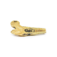
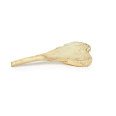
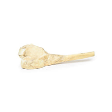
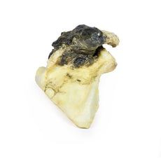
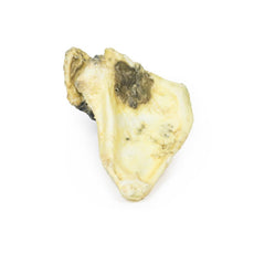
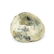
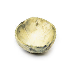
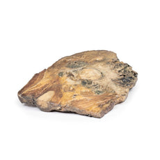
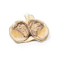
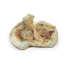
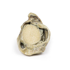
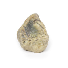
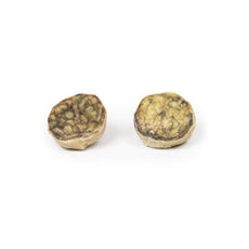
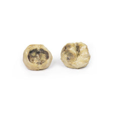
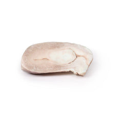
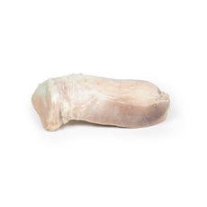
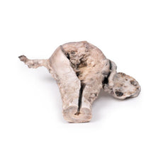
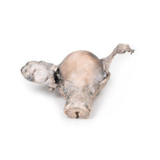
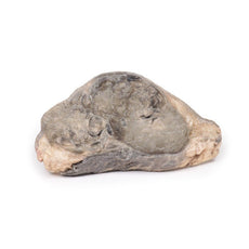
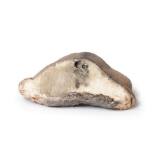
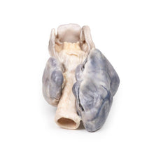
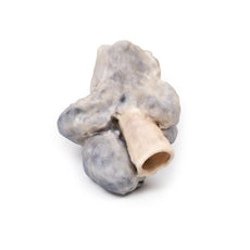
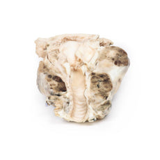
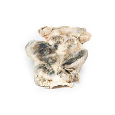
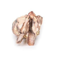
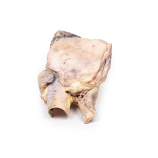
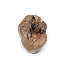
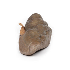
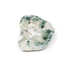
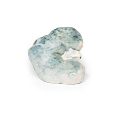
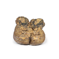
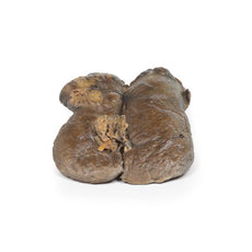
Pathology
This brain specimen has been sliced horizontally. A well-circumscribed 6cm tumour is evident in
between the two frontal lobes. The tumour is compressing the frontal lobes. It has a pinkish cut surface with
some yellow areas indicating necrosis. It was attached to the dura mater anteriorly. This is an example of a
meningioma.
Further Information
Meningiomas are often said to be the most common tumours of the central nervous system
(CNS); however, in fact they arise in the meninges (dura, arachnoid and pia), which are strictly speaking not
part of the CNS per se. They arise from arachnoid cells closely associated with the dura; hence, these tumours
can be associated with the dura or dural folds (falx cerebri and tentorium cerebelli). Meningiomas are
predominantly slow growing benign tumours. Symptoms are determined by the tumour location and the speed of
growth. Symptoms include seizures, change of mental state, vision, hearing- or smell alterations, and symptoms
of increased intracranial pressure. Meningiomas are frequently asymptomatic. Treatment includes observation,
surgery or radiotherapy, depending on the clinical context and tumour morphology.
Meningiomas are rare in children with a median age of 65 years at diagnosis. There is a 3:2 female predominance. Exposure to ionising radiation, including cranial radiotherapy, increases the risk of development meningiomas. The greatest genetic predisposition for development is seen in patients with neurofibromatosis type 2 (NF2). NF2 is an autosomal dominant disease caused by mutations in the NF2 gene on Chromosome 22 leading to multiple tumours associated with the nervous system.
Download:
Handling Guidelines for 3D Printed Models
GTSimulators by Global Technologies
Erler Zimmer Authorized Dealer
These items normal warranty are two years, however the warranty doesn’t cover “wear and tear”. The manufacturer does have 100% quality control on these models.
The models are very detailed and delicate. With normal production machines you cannot realize such details like shown in these models.
The printer used is a color-plastic printer. This is the most suitable printer for these models.
The plastic material is already the best and most suitable material for these prints. (The other option would be a kind of gypsum, but this is way more fragile. You even cannot get them out of the printer without breaking them).The huge advantage of the prints is that they are very realistic as the data is coming from real human specimen. Nothing is shaped or stylized.
The users have to handle these prints with utmost care. They are not made for touching or bending any thin nerves, arteries, vessels etc. The 3D printed models should sit on a table and just rotated at the table.
Related Products


$743.00
$827.00
Free shipping
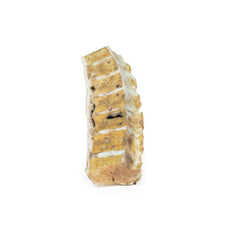
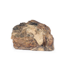
3D Printed Papillary Transitional Cell Carcinoma of the Renal Pelvis
Item # MP2099


by — Item # MP2004
3D Printed Meningioma
$2,893.00
$3,215.00
Add to Cart
Add to Quote
