

Your shopping cart is empty.
3D Printed Pituitary Adenoma

 Handling Guidelines for 3D Printed Models
GTSimulators by Global Technologies
Erler Zimmer Authorized Dealer
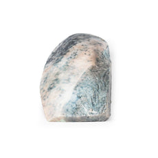
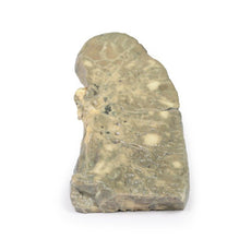
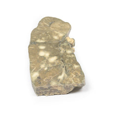
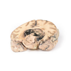


3D Printed Pituitary Adenoma
Item # MP2006
$1,211.00
$1,347.00
You save $136.00
Need an estimate?
Click Add To Quote

Features & Specifications
-
 by
by
A trusted GT partner -
FREE Shipping
U.S. Contiguous States Only -
 3D Printed Model
from a real specimen -
 Gov't pricing
Available upon request
 by
by 
Frequently Bought Together

3D Printed Pituitary Adenoma
Clinical History
A 29-year old male presented with a 22 month history of headaches and blurred vision.
Examination revealed a bi-temporal hemianopia and a left VIth nerve palsy. Skull X-ray showed erosion of most of
the sphenoid body with some dorsum sellae and anterior clinoid process intact. Carotid angiography showed upward
and lateral displacement of the anterior and middle cerebral arteries. Pneumoencephalography (a common imaging
procedure used until the 1970s in which CSF is drained and replaced by air, oxygen or helium that acted as a
contrast medium in X-ray examinations) showed upward displacement of the lateral and third ventricles from
below. A craniotomy was performed but the patient died immediately afterwards.
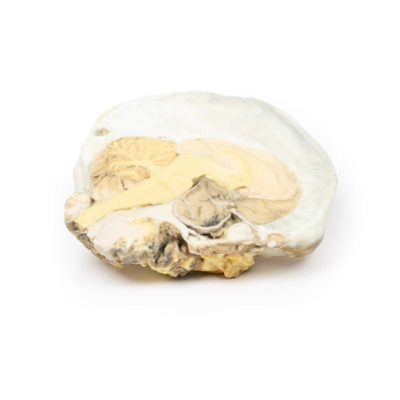
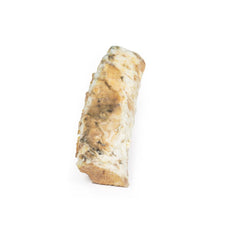
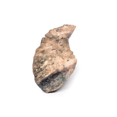
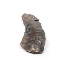
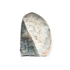
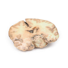
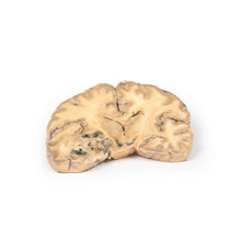
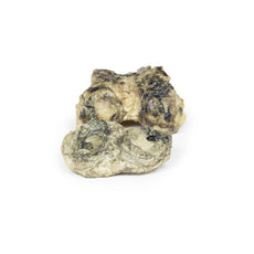
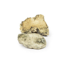
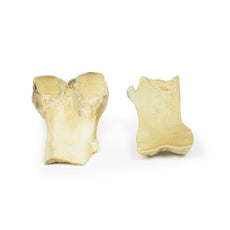
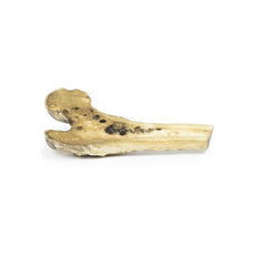
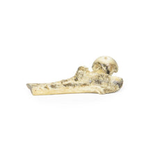
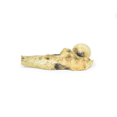
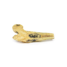
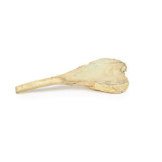
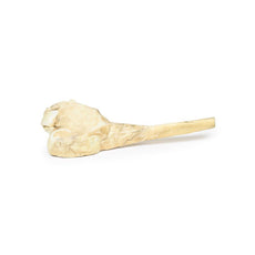
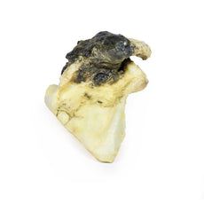
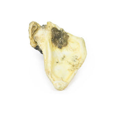
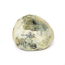
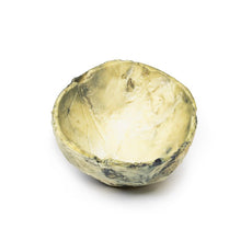
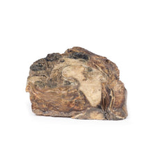
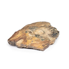
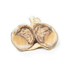
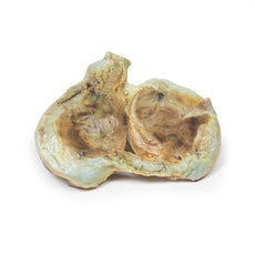
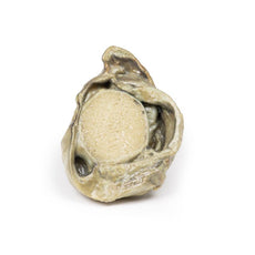
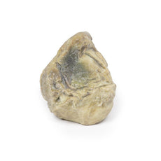
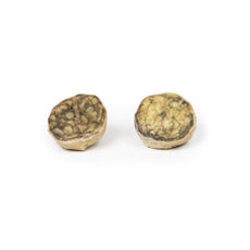
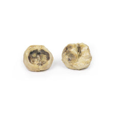
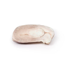
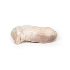
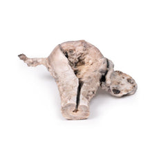
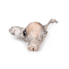
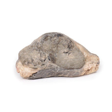
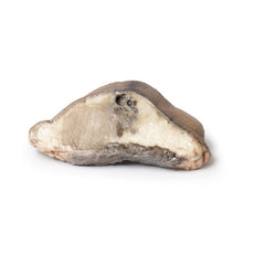
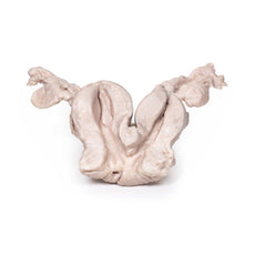
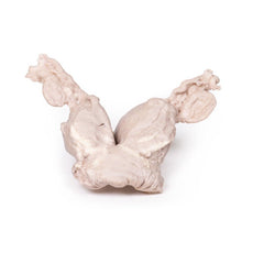
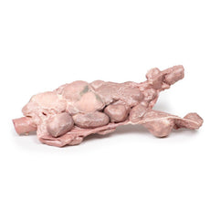
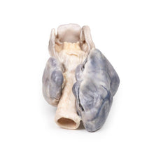
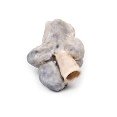
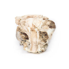
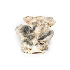
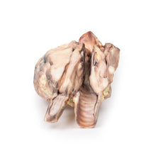
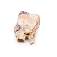
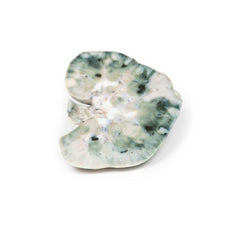
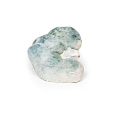
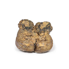
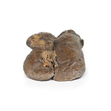
Pathology
The brain specimen is sliced in the sagittal plane to the right of the falx cerebri, which remains
in-situ. The pituitary gland has been completely replaced by a round tumour 4cm in maximum diameter. The tumour
cut surface is pale brown and homogenous (except for an area of haemorrhage superiorly, likely caused by
surgical trauma). The tumour has resulted in upward displacement of the midbrain. Tumour erosion has destroyed
the sphenoid bone; thus, the sella turcica is enlarged (arrow). The optic chiasma is compressed by the tumour.
Histologically, this tumour was a chromophobe adenoma arising from the anterior pituitary.
Further Information
This specimen is from an old case and the investigations used would now be considered
antiquated. Modern investigation would include an initial brain CT followed by an MRI of the brain to further
visualise the pituitary lesion prior to any surgical intervention.
Pituitary adenomas are the most common pituitary tumour and are most commonly found in adults with peak incidence between 35-60 years. Primary carcinoma of the pituitary is very rare, and the pituitary is an uncommon site for metastases. Clinical manifestations of pituitary adenomas are related to local mass effect and tumour function. Local effects include increasing intracranial pressure (headache, nausea and vomiting), sellar expansion, bony erosion and compression of decussating nerve fibres in the optic chiasma, causing bitemporal hemianopia.
Pituitary adenomas can be functioning (i.e. associated with hormone excess) or non-functioning (i.e. without clinical symptoms of hormone excess). About 75% of adenomas are functional: usually secreting prolactin, growth hormone or ACTH. Secretion of TSH, LH and FSH from pituitary adenomas is very rare. Some adenomas can secrete two hormones: growth hormone and prolactin being the most common combination. Non-functional pituitary adenomas come to clinical attention later than those associated with endocrine abnormalities, and they may lead to hypopituitarism due to compression atrophy of the surrounding normal gland.
Download:
Handling Guidelines for 3D Printed Models
GTSimulators by Global Technologies
Erler Zimmer Authorized Dealer
These items normal warranty are two years, however the warranty doesn’t cover “wear and tear”. The manufacturer does have 100% quality control on these models.
The models are very detailed and delicate. With normal production machines you cannot realize such details like shown in these models.
The printer used is a color-plastic printer. This is the most suitable printer for these models.
The plastic material is already the best and most suitable material for these prints. (The other option would be a kind of gypsum, but this is way more fragile. You even cannot get them out of the printer without breaking them).The huge advantage of the prints is that they are very realistic as the data is coming from real human specimen. Nothing is shaped or stylized.
The users have to handle these prints with utmost care. They are not made for touching or bending any thin nerves, arteries, vessels etc. The 3D printed models should sit on a table and just rotated at the table.
Related Products


$743.00
$827.00
Free shipping
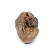
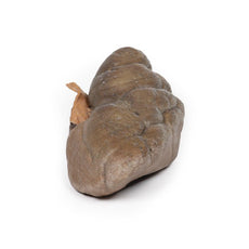
3D Printed Papillary Transitional Cell Carcinoma of the Renal Pelvis
Item # MP2099


by — Item # MP2006
3D Printed Pituitary Adenoma
$1,211.00
$1,347.00
Add to Cart
Add to Quote
