

Your shopping cart is empty.
3D Printed Intracranial Space-Occupying Lesion


 Handling Guidelines for 3D Printed Models
GTSimulators by Global Technologies
Erler Zimmer Authorized Dealer
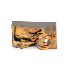
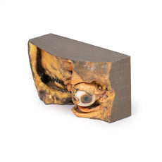
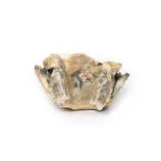
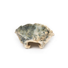
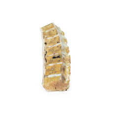
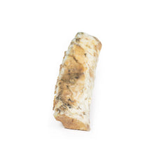

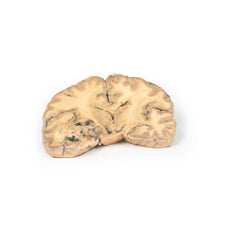
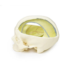

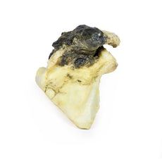
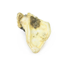
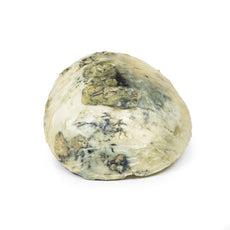
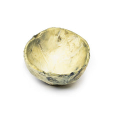


3D Printed Intracranial Space-Occupying Lesion
Item # MP2011
$594.00
$661.00
You save $67.00
Need an estimate?
Click Add To Quote

Features & Specifications
-
 by
by
A trusted GT partner -
 3D Printed Model
from a real specimen -
 Gov't pricing
Available upon request
 by
by 
Frequently Bought Together

3D Printed Intracranial Space-Occupying Lesion
Clinical History
A 56-year-old woman with 6 months of intermittent headache and vomiting was admitted to
hospital comatose after a grand mal seizure, and failed to regain consciousness.
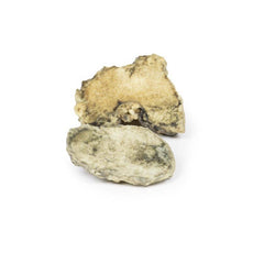
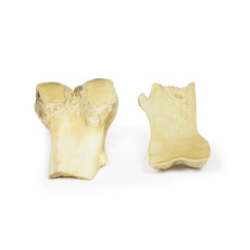
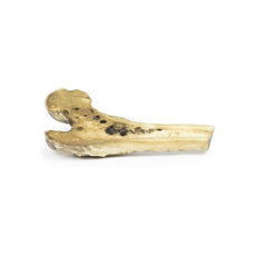
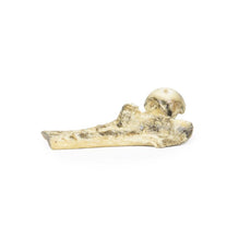
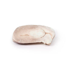
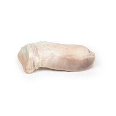
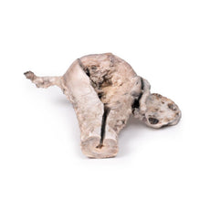
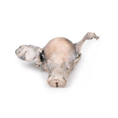
Pathology
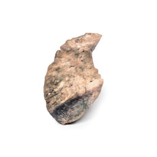
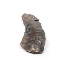
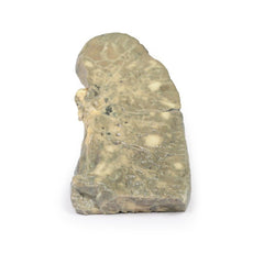
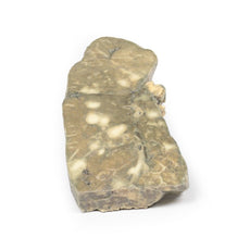
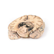
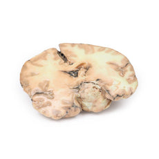
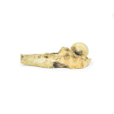
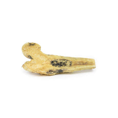
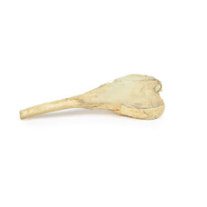
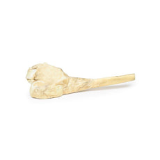
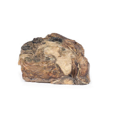
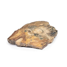
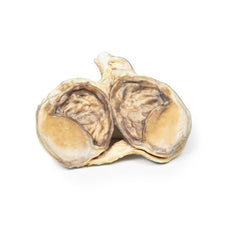
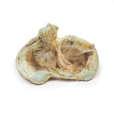
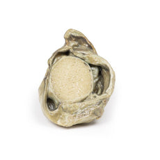
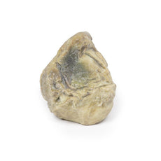
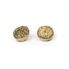
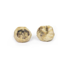
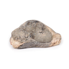
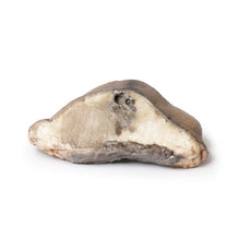
The specimen is a coronal section of a brain. It is evident that the brain has been compressed
laterally and downwards by a right-sided expanding intracranial mass, probably a meningioma. The original mass
is not present. The anterior face shows shift of midline structures with subfalcine herniation* of the cingulate
gyrus. The posterior face (see photo) shows haemorrhage of varying ages within the temporal lobe and the pons,
typical of supratentorial mass lesions. There is also ventricular asymmetry.
*In subfalcine (or cingulate) herniation, the most common type of brain herniation, the innermost part of the frontal lobe is pushed under part of the falx cerebri, between the two hemispheres of the brain.
Further information
Symptoms of a space occupying meningioma in the cranial cavity can be caused by the tumour
mass pressing on the brain, which can lead to atrophy and displacement of brain parenchyma, leading to symptoms
arising from interruptions to cranial nerve functions, blood flow and normal cerebral functions. General
symptoms may include:
Muscle seizures: e.g. Myoclonic (single or multiple muscle twitches, jerks, and/or spasms) or Tonic-clonic (grand
mal: loss of consciousness and body tone, followed by twitching and relaxing muscle contractions, loss of
control of body functions, short period of no breathing and the person may turn a shade of blue, a person may be
sleepy and experience a headache, confusion, weakness, numbness, and sore muscles)
Sensory changes –
alterations in vision, smell, and/or hearing without losing consciousness.
Symptoms and signs may vary with
the location of the tumor.
Handling Guidelines for 3D Printed Models
GTSimulators by Global Technologies
Erler Zimmer Authorized Dealer
These items normal warranty are two years, however the warranty doesn’t cover “wear and tear”. The manufacturer does have 100% quality control on these models.
The models are very detailed and delicate. With normal production machines you cannot realize such details like shown in these models.
The printer used is a color-plastic printer. This is the most suitable printer for these models.
The plastic material is already the best and most suitable material for these prints. (The other option would be a kind of gypsum, but this is way more fragile. You even cannot get them out of the printer without breaking them).The huge advantage of the prints is that they are very realistic as the data is coming from real human specimen. Nothing is shaped or stylized.
The users have to handle these prints with utmost care. They are not made for touching or bending any thin nerves, arteries, vessels etc. The 3D printed models should sit on a table and just rotated at the table.
Related Products

$1,013.00
$1,138.00
Free shipping
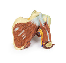
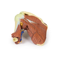
3D Printed Shoulder with deep dissection of the left shoulder
Item # MP1525


by — Item # MP2011
3D Printed Intracranial Space-Occupying Lesion
$594.00
$661.00
Add to Cart
Add to Quote
