Your shopping cart is empty.
3D Printed Intussusception of Small Bowel Due to Metastatic Tumour


 Handling Guidelines for 3D Printed Models
GTSimulators by Global Technologies
Erler Zimmer Authorized Dealer
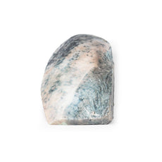
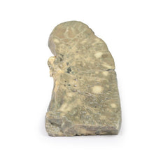
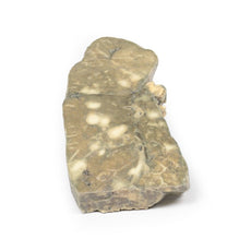
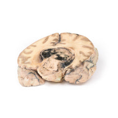


3D Printed Intussusception of Small Bowel Due to Metastatic Tumour
Item # MP2077
$450.00
$501.00
You save $51.00
Need an estimate?
Click Add To Quote

Features & Specifications
-
 by
by
A trusted GT partner -
 3D Printed Model
from a real specimen -
 Gov't pricing
Available upon request
 by
by 
Frequently Bought Together

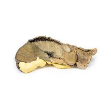
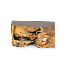
3D Printed Intussusception of Small Bowel Due to Metastatic Tumour
Clinical History
A 66-year-old woman suffered sudden onset of severe colicky central abdominal
pain, somewhat relieved by drawing up her knees. She passed a stool containing mucus and blood (“like redcurrant
jelly”). On examination, there was a mass in the left hypochondrium, which hardened with each spasm of pain. The
specimen was resected at laparotomy.
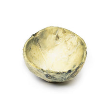
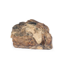
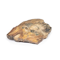
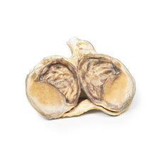
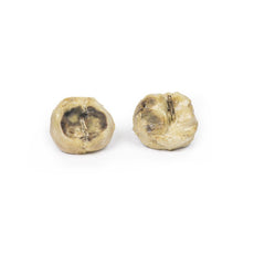
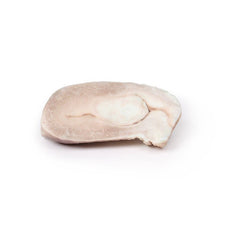
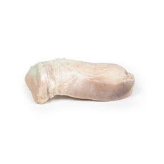
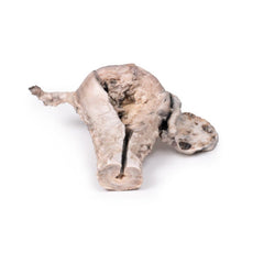
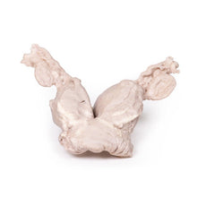
Pathology
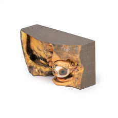
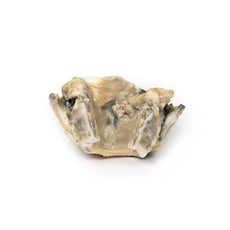
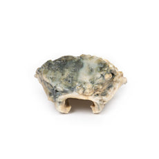
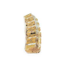
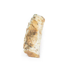
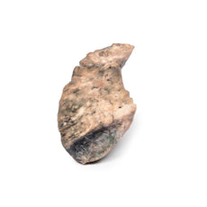
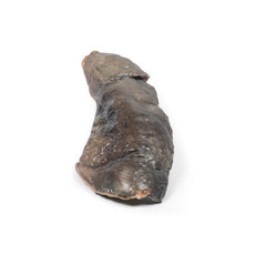
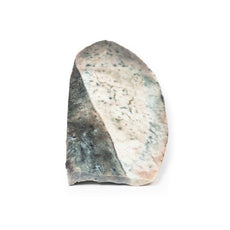
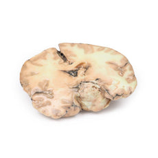
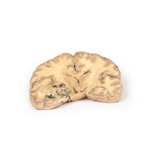
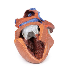
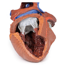
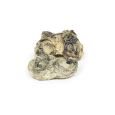
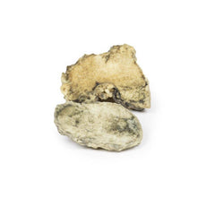
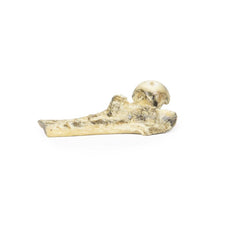
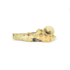
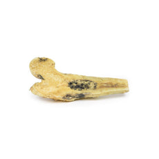
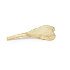
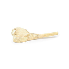
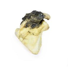
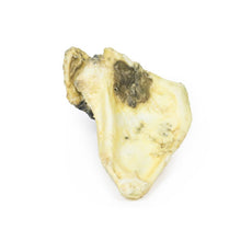
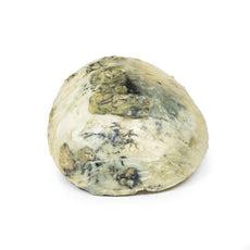
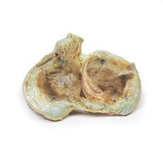
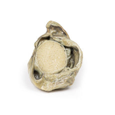
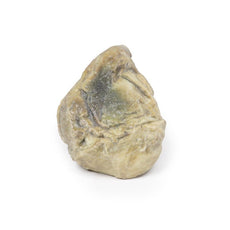
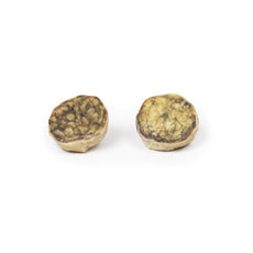
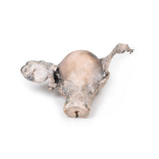
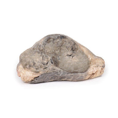
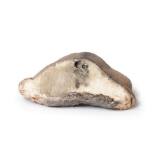
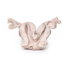
The specimen is a segment of small bowel, approximately 20 cm in length, with
attached mesentery up to 2 cm in width (more evident on the uncut aspect of the specimen). About 5 cm from the
proximal surgical resection margin (which is at the left hand of the specimen), a polypoid tumour 3 cm in diameter
has become invaginated into the lumen of the bowel, and has been propelled distally, forming an intussusception 13
cm in length. The tumour is seen at the apex of the intussusception (near the right hand side of the specimen). The
congestion and exudate seen on the mucosal surface of the intussusception (invaginated portion) are features
considered with early ischaemic necrosis. The histological diagnosis is not recorded in this case; however, the
macroscopic appearance is consistent with a metastatic malignant tumour, although the possibility of a primary
tumour cannot definitely be excluded.
Further Information
Intussusception of the small bowel is most common in children, in whom it is
usually due to invagination of swollen lymphoid tissue (Peyer‘s patches) in the wall of the distal ileum. In adults,
it is rare, causing only between 1 – 5 percent of cases of bowel obstruction. The usual cause a polypoid tumour, as
seen in this specimen, acting as a pathological lead point being pulled forward by peristalsis, and thereby causing
telescoping of the affected portion of bowel distally. Presentation may be of intermittent symptoms of bowel
obstruction and in some cases excruciating pain. Classification of intussusception can be by causal pathology or by
location. Abdominal CT scan will typically demonstrate a typical “target sign” with alternating hyper/hypodense
layers.
Handling Guidelines for 3D Printed Models
GTSimulators by Global Technologies
Erler Zimmer Authorized Dealer
These items normal warranty are two years, however the warranty doesn’t cover “wear and tear”. The manufacturer does have 100% quality control on these models.
The models are very detailed and delicate. With normal production machines you cannot realize such details like shown in these models.
The printer used is a color-plastic printer. This is the most suitable printer for these models.
The plastic material is already the best and most suitable material for these prints. (The other option would be a kind of gypsum, but this is way more fragile. You even cannot get them out of the printer without breaking them).The huge advantage of the prints is that they are very realistic as the data is coming from real human specimen. Nothing is shaped or stylized.
The users have to handle these prints with utmost care. They are not made for touching or bending any thin nerves, arteries, vessels etc. The 3D printed models should sit on a table and just rotated at the table.
Related Products

$1,013.00
$1,138.00
Free shipping
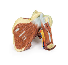
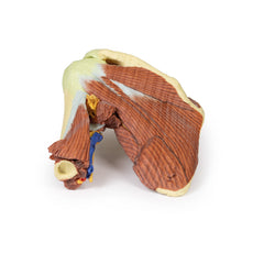
3D Printed Shoulder with deep dissection of the left shoulder
Item # MP1525


by — Item # MP2077
3D Printed Intussusception of Small Bowel Due to Metastatic Tumour
$450.00
$501.00
Add to Cart
Add to Quote