

Your shopping cart is empty.
3D Printed Metastatic Adenocarcinoma in the Brain

 Handling Guidelines for 3D Printed Models
GTSimulators by Global Technologies
Erler Zimmer Authorized Dealer
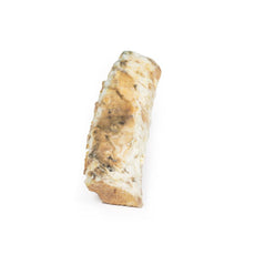
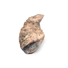
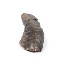
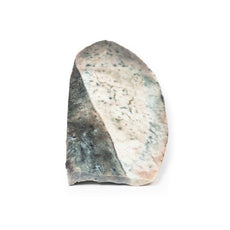
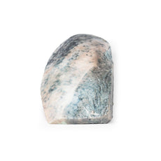
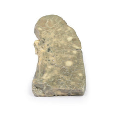
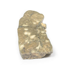
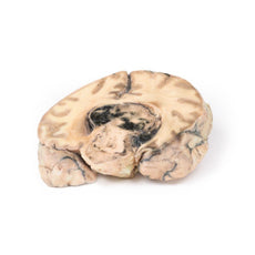

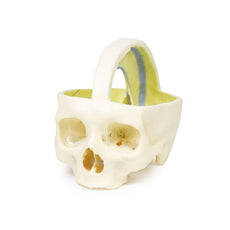
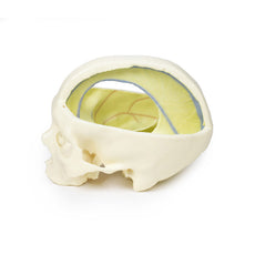

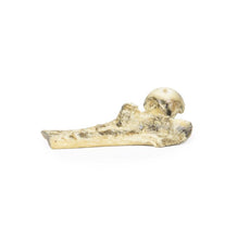
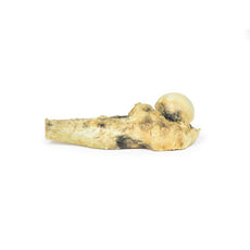
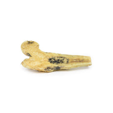
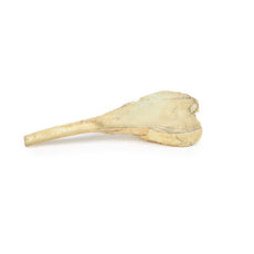
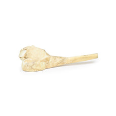
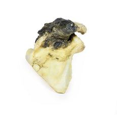
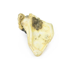
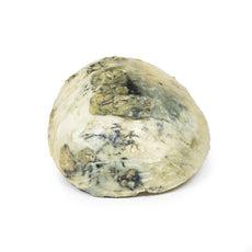


3D Printed Metastatic Adenocarcinoma in the Brain
Item # MP2007
$354.00
$395.00
You save $41.00
Need an estimate?
Click Add To Quote

Features & Specifications
-
 by
by
A trusted GT partner -
 3D Printed Model
from a real specimen -
 Gov't pricing
Available upon request
 by
by 
Frequently Bought Together

3D Printed Metastatic Adenocarcinoma in the Brain
Clinical History
A 56-year old male underwent a total gastrectomy and splenectomy for gastric
adenocarcinoma. Over a period of two months he developed a progressively unsteady gait,
increasing weakness of his left hand and frontal headaches associated with nausea and vomiting.
Imaging revealed a lesion in the right frontal lobe. He underwent a craniotomy with resection of
the lesion, which was confirmed metastatic gastric adenocarcinoma. He experienced gradual
increasing symptoms as well as jaundice, deteriorating consciousness and papilloedema from
increased intracranial pressure. Repeat imaging revealed recurrence of the right frontal
metastatic lesion as well as liver metastases. The patient died 9 months after his initial
gastrectomy surgery.
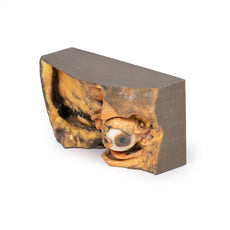
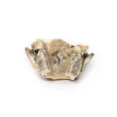
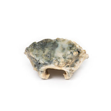
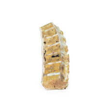
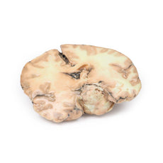
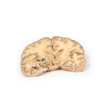
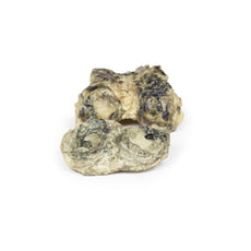
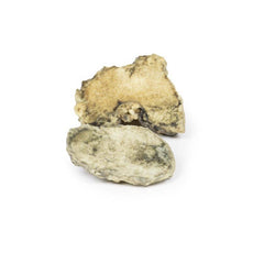
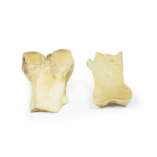
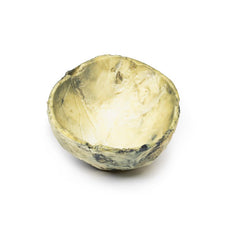
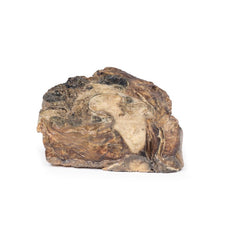
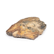
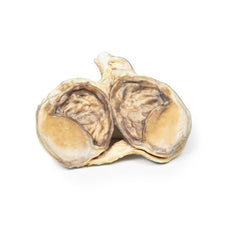
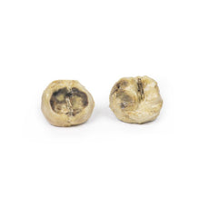
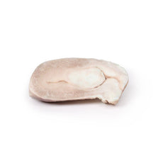
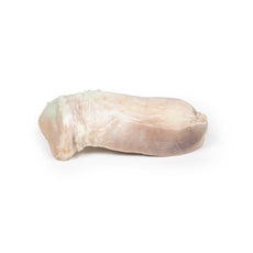
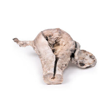
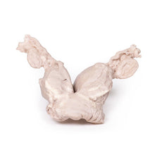
Pathology
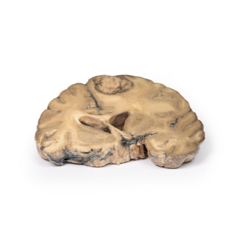
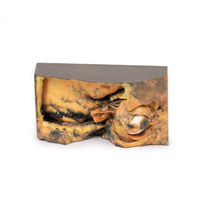
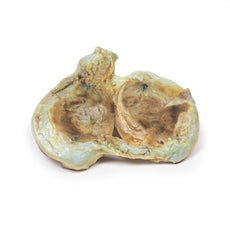
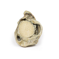
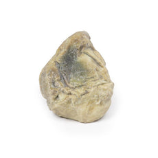
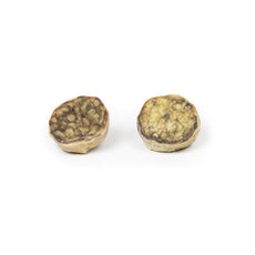
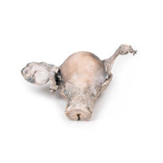
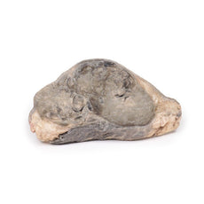
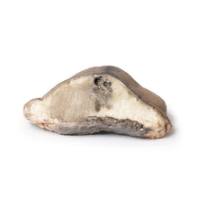
This brain specimen is cut in the coronal plane. A circumscribed, variegated,
pink-grey tumour is evident in the right frontal lobe. The tumour is involving the grey and
white matter. Compression of the right lateral ventricle by the lesion is apparent with shift of
the midline structures also seen.
Further Information
Stomach cancer is one of the most common causes of cancer-related death
worldwide. Risk factors include male gender, diet, smoking and chronic Helicobacter pylori
infection. The most common sites for metastases of gastric adenocarcinoma are the liver,
peritoneum, lungs and bones. Brain metastases are rare, occurring in <1% of cases. Isolated
brain metastases are very uncommon with them being more commonly seen in disseminated disease
and associated with a poor prognosis. Palliative treatment may include surgery, radiotherapy,
steroid, chemotherapy or a combination thereof.
Handling Guidelines for 3D Printed Models
GTSimulators by Global Technologies
Erler Zimmer Authorized Dealer
These items normal warranty are two years, however the warranty doesn’t cover “wear and tear”. The manufacturer does have 100% quality control on these models.
The models are very detailed and delicate. With normal production machines you cannot realize such details like shown in these models.
The printer used is a color-plastic printer. This is the most suitable printer for these models.
The plastic material is already the best and most suitable material for these prints. (The other option would be a kind of gypsum, but this is way more fragile. You even cannot get them out of the printer without breaking them).The huge advantage of the prints is that they are very realistic as the data is coming from real human specimen. Nothing is shaped or stylized.
The users have to handle these prints with utmost care. They are not made for touching or bending any thin nerves, arteries, vessels etc. The 3D printed models should sit on a table and just rotated at the table.
Related Products

$1,013.00
$1,138.00
Free shipping
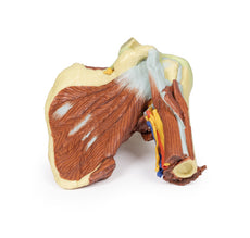
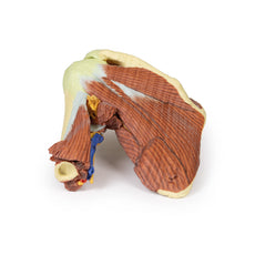
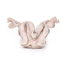
3D Printed Shoulder with deep dissection of the left shoulder
Item # MP1525


by — Item # MP2007
3D Printed Metastatic Adenocarcinoma in the Brain
$354.00
$395.00
Add to Cart
Add to Quote
