

Your shopping cart is empty.
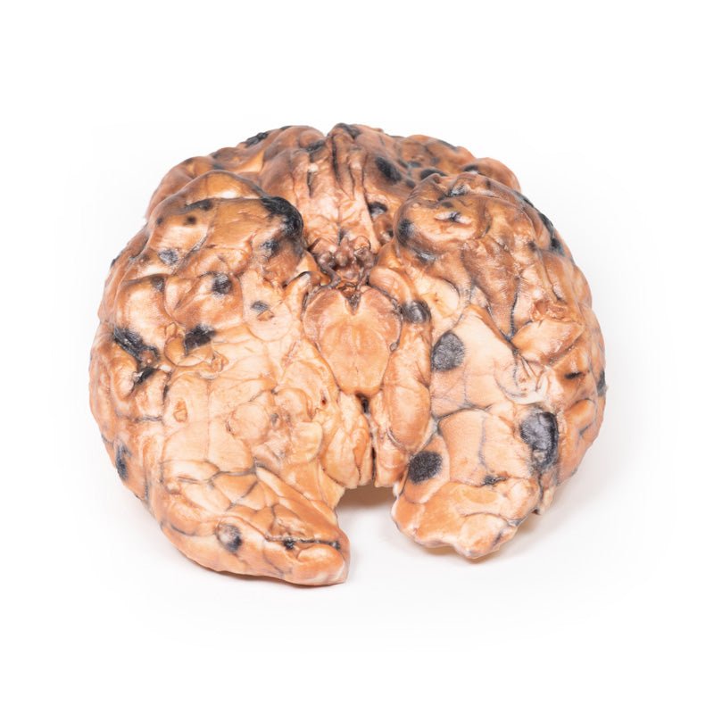
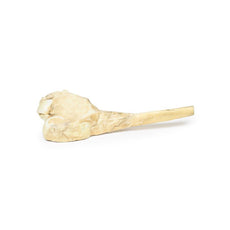
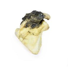
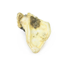
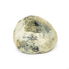
3D Printed Metastatic Melanoma

 Handling Guidelines for 3D Printed Models
GTSimulators by Global Technologies
Erler Zimmer Authorized Dealer
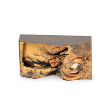
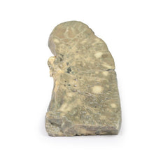
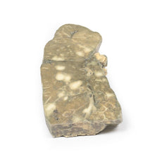
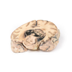

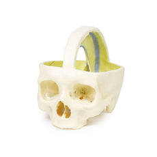
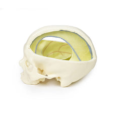


3D Printed Metastatic Melanoma
Item # MP2018
$986.00
$1,097.00
You save $111.00
Need an estimate?
Click Add To Quote

Features & Specifications
-
 by
by
A trusted GT partner -
FREE Shipping
U.S. Contiguous States Only -
 3D Printed Model
from a real specimen -
 Gov't pricing
Available upon request
 by
by 
Frequently Bought Together

3D Printed Metastatic Melanoma
Clinical History
In the 1970s, a 31-year-old woman presented with severe headache and diplopia on a background
of having a pigmented skin lesion (diagnosed as an invasive skin melanoma) removed from her neck 8 months
earlier. Clinical examination revealed no abnormality, and following discharge the patient was later re-admitted
with persistent vomiting. Her condition deteriorated and she died.
Pathology
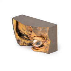
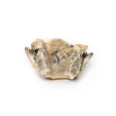
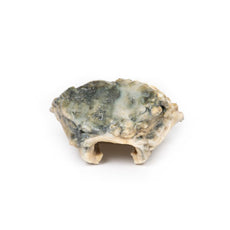
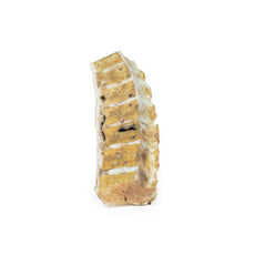
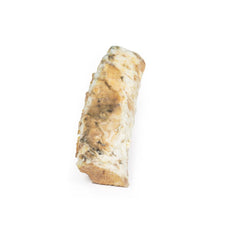
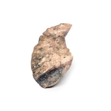
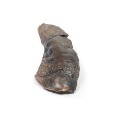
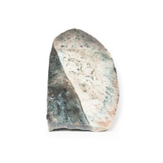
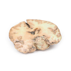
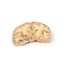
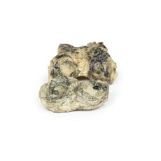
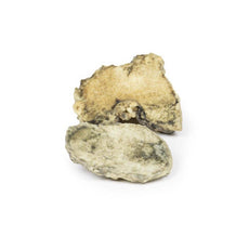
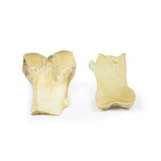
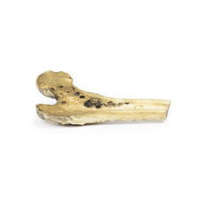
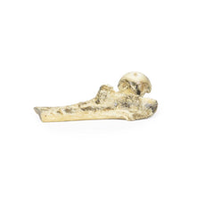
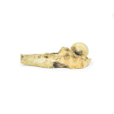
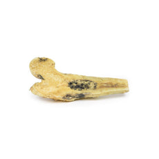
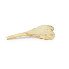
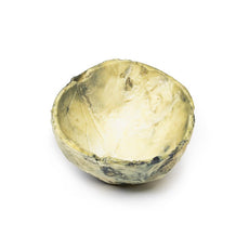
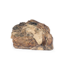
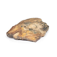
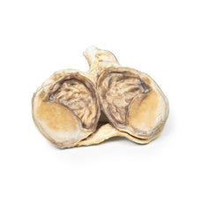
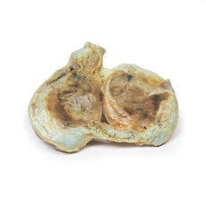
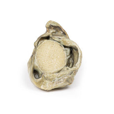
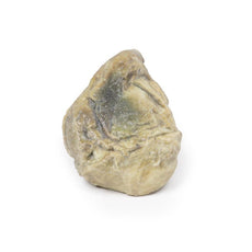
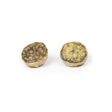
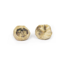
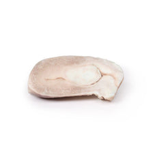
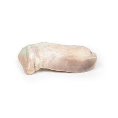
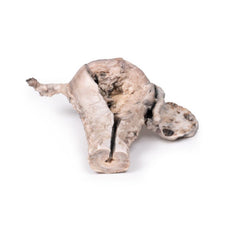
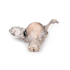
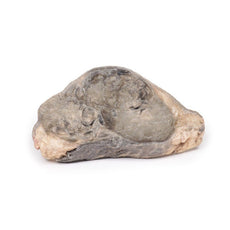
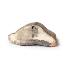
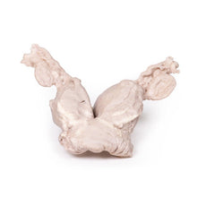
This specimen demonstrates widespread intracerebral melanoma metastases. The inferior surface is
characterised by many elevated dark nodules up to 1.5 cm in diameter. Similar lesions are present on the cut
superior surface where it is seen that these secondary melanotic deposits are confined exclusively to the grey
matter. The tumour deposits are not encapsulated and are invading the cortex. Some necrosis and haemorrhage is
present.
Further information
Of all patients who have metastatic disease to the brain, 10% are from skin melanoma. Risk
increases with age over 60 years, male gender, disease duration and more advanced tumour/metastatic stage. BRAF
and NRAS mutations, expression of CCR4 receptors on tumour cells, and activation of the PI3K pathway are all
risk factors for the development of cerebral metastasis. 80% of melanoma brain metastases are supratentorial.
Presentation is often with headache, neurologic deficits and/or seizures. Furthermore, these lesions are at risk
of spontaneous haemorrhage. Modern diagnosis is based on neuroimaging and often histology of a stereotactic
brain biopsy, if no previous diagnosis has been made. Treatment includes stereotactic radiosurgery (SRS),
radiotherapy and/or systemic therapy with “checkpoint inhibitor immunotherapy” or targeted treatments. This has
improved median survival upto 11 months in recent years.\
Handling Guidelines for 3D Printed Models
GTSimulators by Global Technologies
Erler Zimmer Authorized Dealer
These items normal warranty are two years, however the warranty doesn’t cover “wear and tear”. The manufacturer does have 100% quality control on these models.
The models are very detailed and delicate. With normal production machines you cannot realize such details like shown in these models.
The printer used is a color-plastic printer. This is the most suitable printer for these models.
The plastic material is already the best and most suitable material for these prints. (The other option would be a kind of gypsum, but this is way more fragile. You even cannot get them out of the printer without breaking them).The huge advantage of the prints is that they are very realistic as the data is coming from real human specimen. Nothing is shaped or stylized.
The users have to handle these prints with utmost care. They are not made for touching or bending any thin nerves, arteries, vessels etc. The 3D printed models should sit on a table and just rotated at the table.
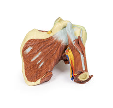
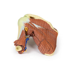
Related Products

$1,013.00
$1,138.00
Free shipping
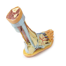
3D Printed Shoulder with deep dissection of the left shoulder
Item # MP1525


by — Item # MP2018
3D Printed Metastatic Melanoma
$986.00
$1,097.00
Add to Cart
Add to Quote
