

Your shopping cart is empty.
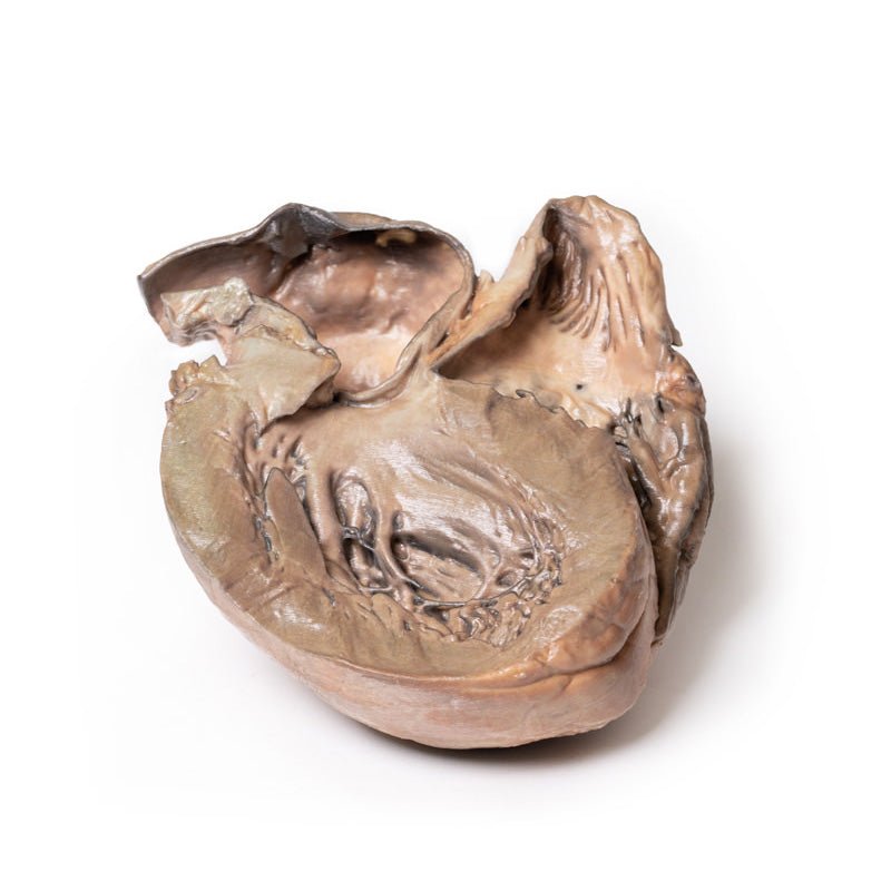
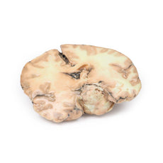
3D Printed Ruptured Thoracic Aortic Aneurysm

 Handling Guidelines for 3D Printed Models
GTSimulators by Global Technologies
Erler Zimmer Authorized Dealer
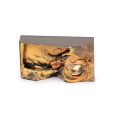
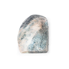
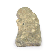
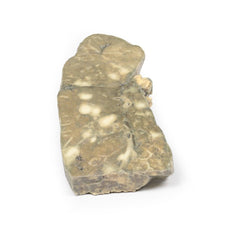
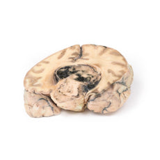


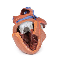
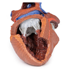
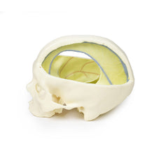


3D Printed Ruptured Thoracic Aortic Aneurysm
Item # MP2043
$1,258.00
$1,399.00
You save $141.00
Need an estimate?
Click Add To Quote

Features & Specifications
-
 by
by
A trusted GT partner -
FREE Shipping
U.S. Contiguous States Only -
 3D Printed Model
from a real specimen -
 Gov't pricing
Available upon request
 by
by 
Frequently Bought Together

3D Printed Ruptured Thoracic Aortic Aneurysm
Clinical History
No clinical details are available for this specimen.
Pathology
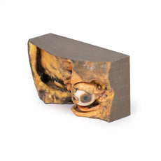
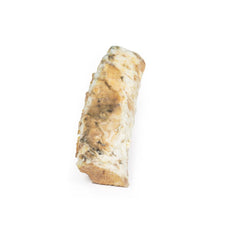
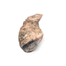
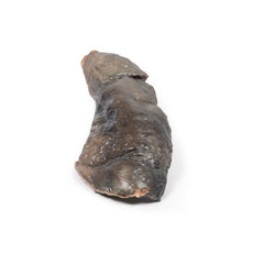
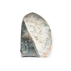
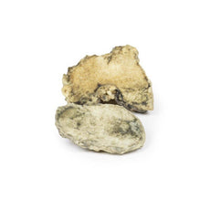
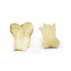
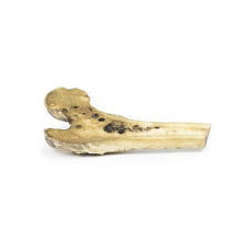
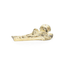
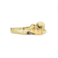
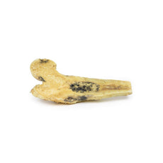
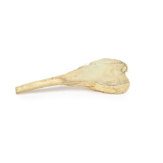
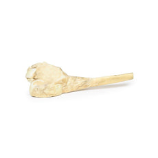
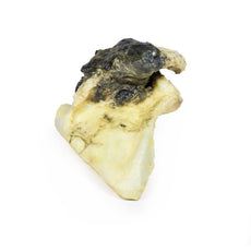
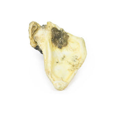
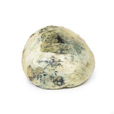
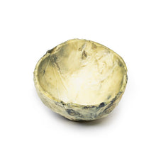
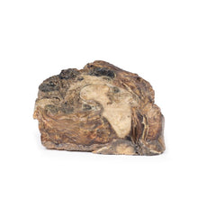
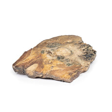
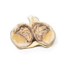
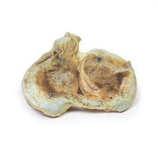
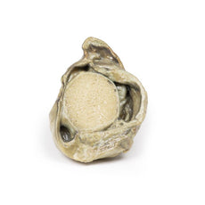
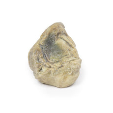
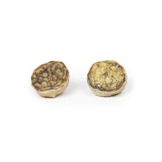
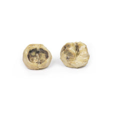
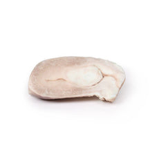
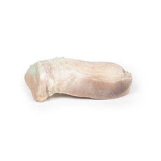
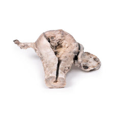
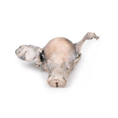
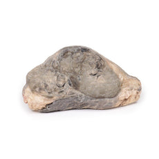
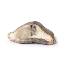
The heart displays both ventricles from the posterior aspect. There is a prominent saccular dilatation
of the thoracic ascending aorta, which shows several atherosclerotic plaques and posteriorly is seen to be ruptured
(identified by the dark staining). Both ventricles are hypertrophied. The coronary arteries together with the aortic
and tricuspid valves are normal. This is an example of a ruptured aneurysm of the ascending aorta.
Further information
The dilation of the ascending aorta is a common incidental finding on transthoracic
echocardiography performed for unrelated indications.
The thoracic aorta is divided into 3 parts: ascending, arch
and descending. The ascending aorta originates beyond the aortic valve and ends right before the innominate artery
(brachiocephalic trunk). It is approximately 5 cm long and is composed of two distinct segments. The lower segment,
known as the aortic root, encompasses the coronary sinuses and sinotubular junction (STJ). The upper segment, known
as the tubular ascending aorta, begins at the STJ and extends to the aortic arch (innominate artery). More than 50%
of thoracic aortic aneurysms are localized to the ascending aorta, which may affect either the aortic root or
tubular aortic segment.
An aneurysm is defined as a localized dilation of the aorta that is more than 50% of
predicted (ratio of observed to expected diameter = 1.5). Aneurysm should be distinguished from ectasia, which
represents a diffuse dilation of the aorta less than 50% of normal aorta diameter. The incidence of ascending
thoracic aortic aneurysms is estimated to be around 10 per 100,000 person-years[1].
Reference
1. Saliba et al. (2015). Int J Cardiol Heart Vasc. 6: 91–100.
Handling Guidelines for 3D Printed Models
GTSimulators by Global Technologies
Erler Zimmer Authorized Dealer
These items normal warranty are two years, however the warranty doesn’t cover “wear and tear”. The manufacturer does have 100% quality control on these models.
The models are very detailed and delicate. With normal production machines you cannot realize such details like shown in these models.
The printer used is a color-plastic printer. This is the most suitable printer for these models.
The plastic material is already the best and most suitable material for these prints. (The other option would be a kind of gypsum, but this is way more fragile. You even cannot get them out of the printer without breaking them).The huge advantage of the prints is that they are very realistic as the data is coming from real human specimen. Nothing is shaped or stylized.
The users have to handle these prints with utmost care. They are not made for touching or bending any thin nerves, arteries, vessels etc. The 3D printed models should sit on a table and just rotated at the table.
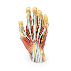
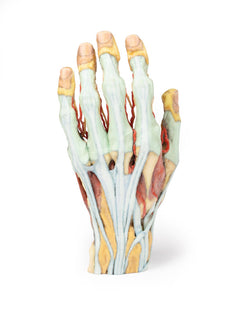
Related Products

$1,013.00
$1,138.00
Free shipping
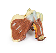
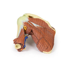
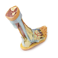
3D Printed Shoulder with deep dissection of the left shoulder
Item # MP1525


by — Item # MP2043
3D Printed Ruptured Thoracic Aortic Aneurysm
$1,258.00
$1,399.00
Add to Cart
Add to Quote
