

Your shopping cart is empty.
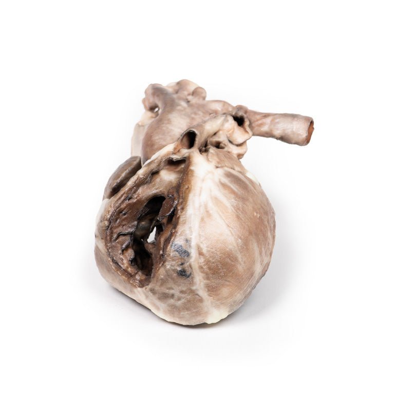
3D Printed Tetralogy of Fallot

 Handling Guidelines for 3D Printed Models
GTSimulators by Global Technologies
Erler Zimmer Authorized Dealer
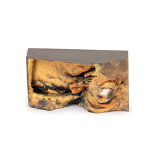
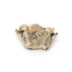
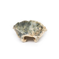
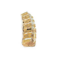
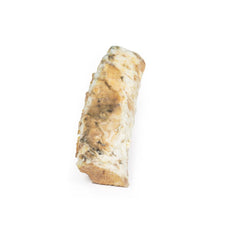
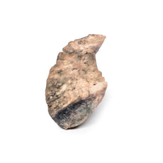
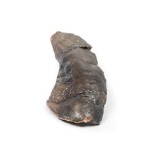
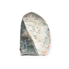
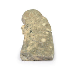
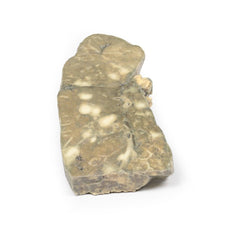
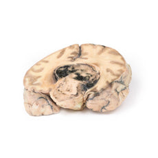


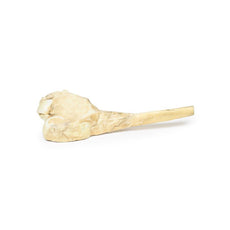
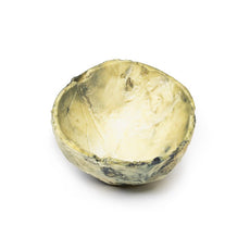
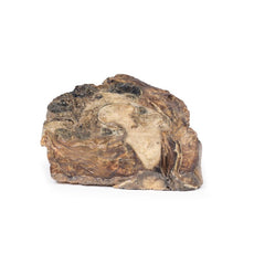
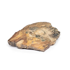
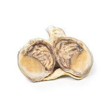


3D Printed Tetralogy of Fallot
Item # MP2037
$466.00
$519.00
You save $53.00
Need an estimate?
Click Add To Quote

Features & Specifications
-
 by
by
A trusted GT partner -
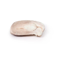
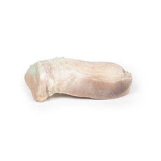
 3D Printed Model
from a real specimen -
 Gov't pricing
Available upon request
 by
by 
Frequently Bought Together

3D Printed Tetralogy of Fallot
Clinical History
A 21-month old boy was admitted with a history of exhaustion and exertional dyspnoea for the
previous 2 to 3 months. During this time there had been several attacks of acute dyspnoea each lasting up to two
minutes. Examination revealed central cyanosis, mild finger clubbing, and a harsh systolic bruit maximal at the
left sternal edge. Cardiac catheterisation led to a diagnosis of Fallot‘s tetralogy and severe pulmonary oedema.
A surgical correction was performed (Willis-Potts anastomosis between the aorta and the origin of the left
pulmonary artery). The child developed acute dyspnoea and left lobar consolidation 12 hours post-operatively and
died despite treatment.
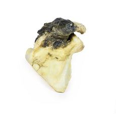
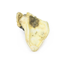
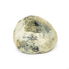
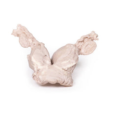
Pathology
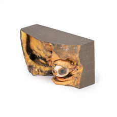
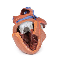
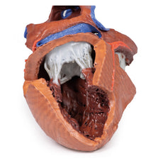
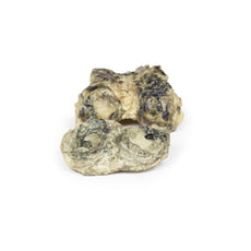
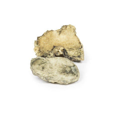
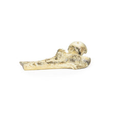
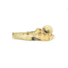
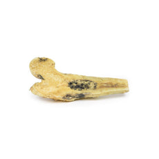
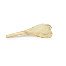
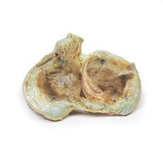
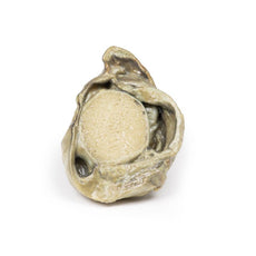
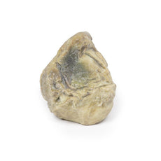
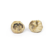
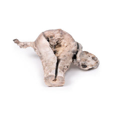
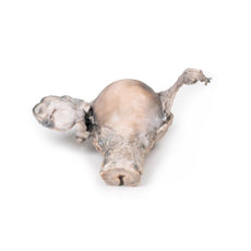
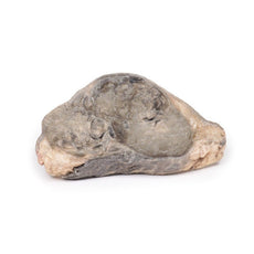
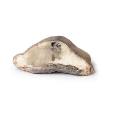
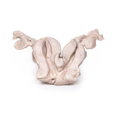
The child‘s heart is viewed from the anterior aspect. The anterior wall of the right ventricle has
been excised to reveal prominent right ventricular hypertrophy and a narrowed pulmonary outflow tract. The
pulmonary valve ring is also small, with a bicuspid stenosed valve. There is a patch of endocardial fibrosis in
the outflow tract below the pulmonary valve. The origin of the aorta overlies a high ventricular septal defect.
A probe could be passes into the aorta from the hypertrophied right ventricle. The further probe was able to be
passed from the narrowed pulmonary trunk into a dilated, thin-walled left pulmonary artery and through the
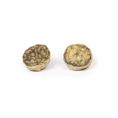
surgical anastomosis into the descending aorta. Examination of the posterior aspect of the specimen reveals an
opened right atrium and left ventricle. When viewed from the right side of the heart, there is a large atrial
septal defect (ASD), 8 mm in diameter at the site of the foramen ovale (large arrow). Another tiny ASD (small
arrow) 3 mm in diameter is present posterior to the upper border of the large ASD. Note that the wall of the
left ventricle is slightly thinner than the wall of the right ventricle.
Further Information
The four features of tetralogy of Fallot are: 1. Ventricular septal defect (VSD); 2. An
aorta that straddles the VSD with the latter communicating with both ventricles (over-riding aorta) instead of
just the left ventricle; 3. Pulmonary stenosis or obstruction of the right ventricular overflow tract; 4. Right
ventricular hypertrophy. This condition usually causes cyanosis early in life. Its severity depends on the
degree of pulmonary outflow obstruction, which determines whether there is a left-to-right, or right-to-left
shunt. In some patients, pulmonary blood flow is increased due to the presence of a patent ductus arteriosus.
Patients with this condition may survive untreated into adult life, and a few may reach middle age. However,
surgical correction is now possible and is desirable, as the disorder is ultimately fatal. Sometimes additional
cardiac abnormalities may be present. (e.g. atrial septal defect, as was found in this case).
In most cases
of tetralogy of Fallot, the cause is not known although in some patients, genetic factors play a role. For
example, the condition is more common in patients with Down syndrome (Trisomy 21; in association with AV canal
defects) or DiGeorge syndrome (22q11 deletion).
Handling Guidelines for 3D Printed Models
GTSimulators by Global Technologies
Erler Zimmer Authorized Dealer
These items normal warranty are two years, however the warranty doesn’t cover “wear and tear”. The manufacturer does have 100% quality control on these models.
The models are very detailed and delicate. With normal production machines you cannot realize such details like shown in these models.
The printer used is a color-plastic printer. This is the most suitable printer for these models.
The plastic material is already the best and most suitable material for these prints. (The other option would be a kind of gypsum, but this is way more fragile. You even cannot get them out of the printer without breaking them).The huge advantage of the prints is that they are very realistic as the data is coming from real human specimen. Nothing is shaped or stylized.
The users have to handle these prints with utmost care. They are not made for touching or bending any thin nerves, arteries, vessels etc. The 3D printed models should sit on a table and just rotated at the table.
Related Products

$1,013.00
$1,138.00
Free shipping
3D Printed Shoulder with deep dissection of the left shoulder
Item # MP1525


by — Item # MP2037
3D Printed Tetralogy of Fallot
$466.00
$519.00
Add to Cart
Add to Quote
