Your shopping cart is empty.
3D Printed Abdomen with Bilateral Hernias

 Handling Guidelines for 3D Printed Models
GTSimulators by Global Technologies
Erler Zimmer Authorized Dealer


3D Printed Abdomen with Bilateral Hernias
Item # MP1130
$11,589.00
$12,877.00
You save $1,288.00
Need an estimate?
Click Add To Quote

Features & Specifications
-
 by
by
A trusted GT partner -
FREE Shipping
U.S. Contiguous States Only -
 3D Printed Model
from a real specimen -
 Gov't pricing
Available upon request
 by
by 
Frequently Bought Together

3D Printed Abdomen with Bilateral Hernias
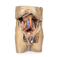
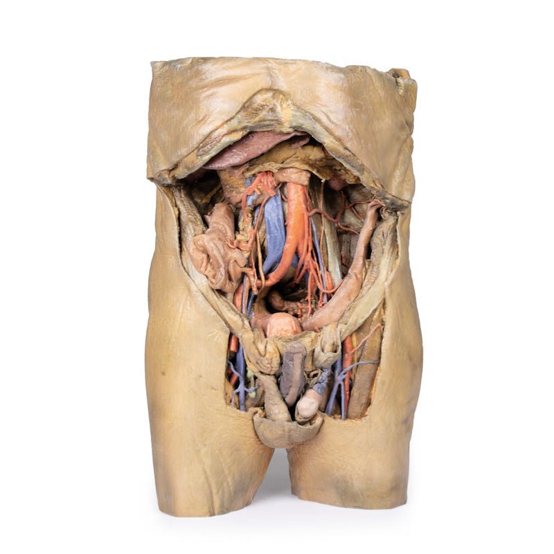
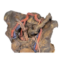
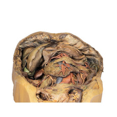
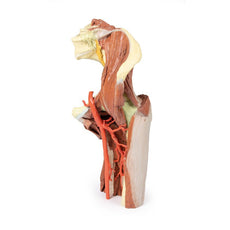
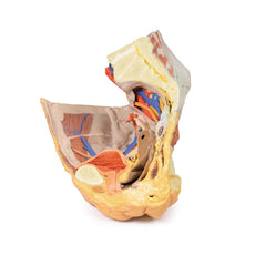
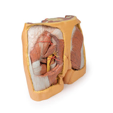
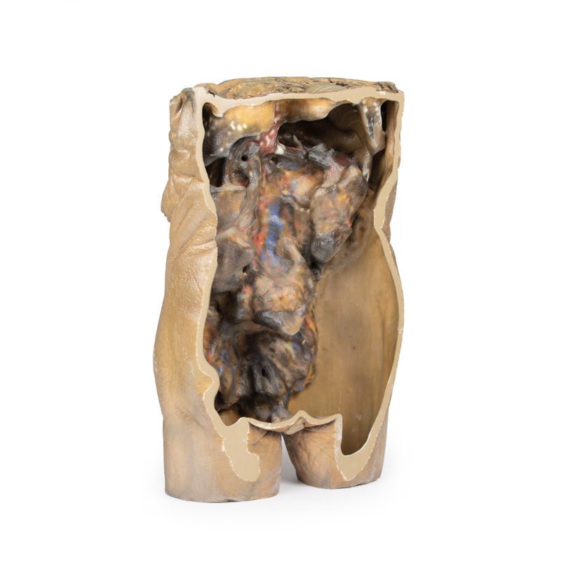
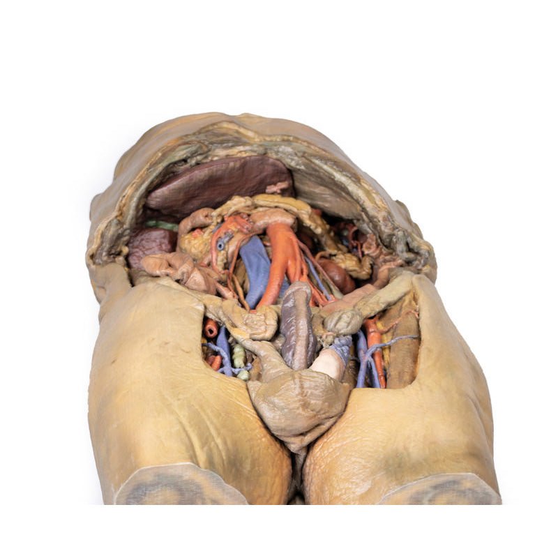
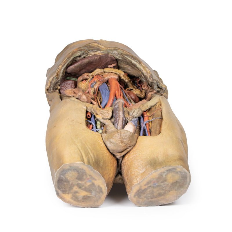
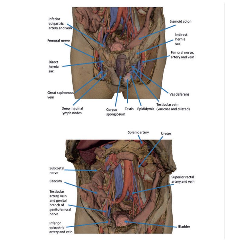
This 3D model represents one of the largest and most complex in the series, consisting of a partial torso from the diaphragm to the proximal thigh with a complete abdominal cavity preserving varying levels of dissection. This 3D model also records the rare, simultaneous occurrence of indirect and direct inguinal hernias allowing for a consideration of the anatomical underpinnings for both conditions. Given the scale of the dissection this 3D model description is divided into discrete parts based on views and regions.
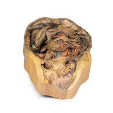
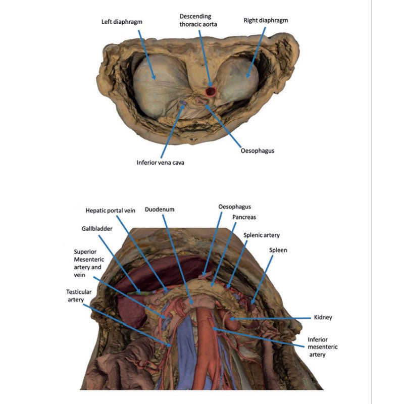
The diaphragm
On the superior aspect of the model the diaphragm is preserved, and while slightly distorted due
to removal of the thoracic ribs through dissection, both domes and costodiaphragmatic recesses can be
appreciated. The fibrous pericardium is present on the superior surface of the central tendon, with the terminal
part of the inferior vena cava visible in the caval foramen. Just lateral to caval foramen is the oesophagus
within the oesophageal hiatus, and then the descending thoracic aorta approaching the aortic hiatus just ventral
to the thoracic vertebrae.
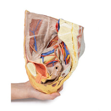
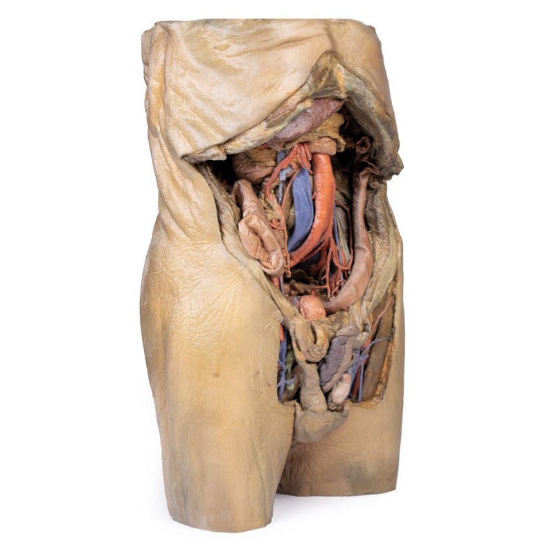
The epigastric and hypochondriac regions
Within the abdomen, the anterior abdominal wall, greater omentum, and
much of the gastrointestinal tract has been removed alongside the parietal peritoneum over the posterior
abdominal wall to expose retroperitoneal organs and structures. In the superior abdomen, the terminal portion of
the oesophagus has been retained and can be seen entering the cavity just lateral to the left lobe of the liver.
The removal of the stomach has exposed the extent of the pancreas from the head (positioned within the arc of
the duodenum) to the tail extending to the capsule of the spleen preserved in the left hypochondrium. Superior
to the pancreas, the splenic artery and common hepatic arteries can just be observed spanning across the narrow
space between the pancreas, diaphragm and liver. The splenic follows its archetypical ‘tortuous route’
towards the spleen, and strongly divides prior to reaching the hilum (and adjacent to the splenic vein). The
common hepatic can be seen dividing into the gastroduodenal (visible again as a cut vessel just inferior to the
duodenum) and giving off the right gastric artery; these vessels lie superficial relative to the hepatic portal
vein. The superior mesenteric artery and vein can be seen passing anteriorly near the head of the pancreas and
horizontal part of the duodenum, and the retained ileocolic artery can be traced to the caecum of the large
intestine in the lower right quadrant of the abdomen. The inferior mesenteric vein can be, in part, appreciated
arising from the retained superior rectal vein ascending from the undissected true pelvis and spanning across
the superficial aspect of the descending thoracic aorta.
Inferior to the liver the gallbladder can be viewed just between the right and left anatomical lobes. On the left, the passage of the renal artery and vein can be seen just deep to the pancreas, and the ureters can be observed descending from the partially exposed kidney across the superficial surface of the exposed psoas major and minor muscles.
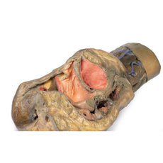
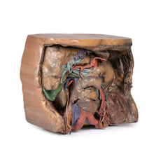
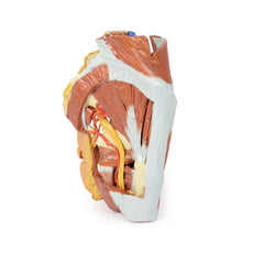
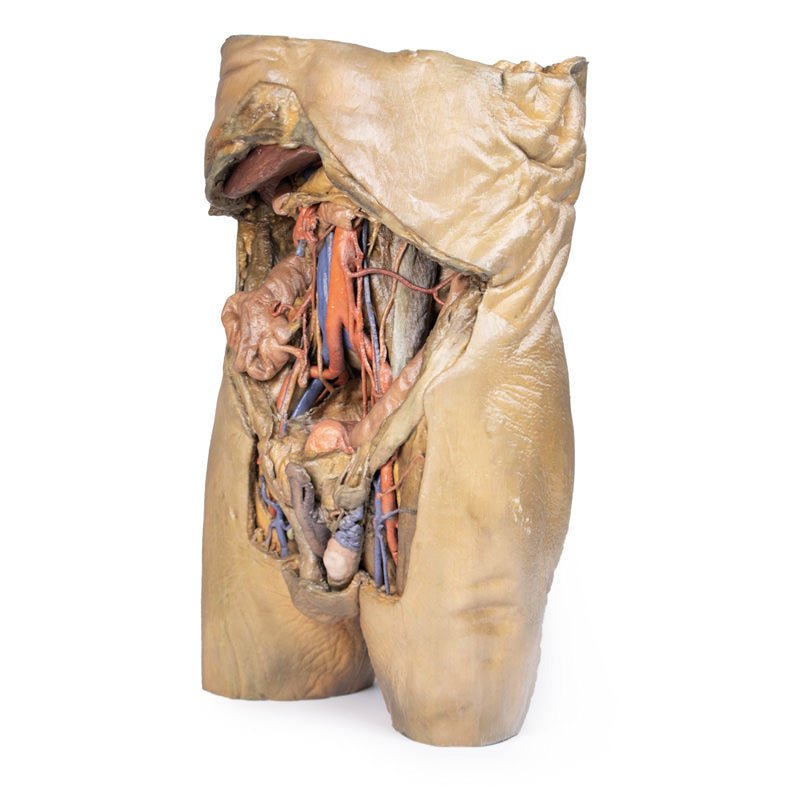
The umbilical and lumbar regions
Most of the organs occupying the umbilical and lumbar regions of the abdomen
have been removed in order to expose structures in the posterior abdominal wall. In the midline, the descending
abdominal aorta and inferior vena cava dominate the region, with the testicular arteries and veins isolated and
traceable towards the inguinal regions. Two right lumbar arteries are visible arising from the aorta, and
despite removal of the mesenteries and most of the colon the inferior mesenteric artery can be seen giving rise
to the left colic, sigmoid and superior rectal arteries. On the right side of the specimen inferior to the
kidney, the subcostal, iliohypogastric and ilioingual nerves are exposed alongside the circumflex iliac artery.
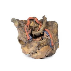
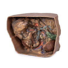
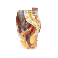
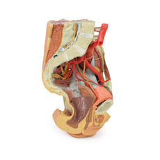
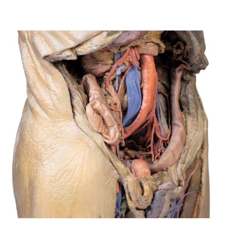
The hypogastrium and iliac regions
In the midline, the bifurcation of the descending abdominal aorta into the
common iliacs (and subsequent division into the internal and external iliacs) can be observed deep to some of
the overlying structures (e.g., testicular vessels, ureters) noted previously. On the right side, the obturator
artery can be seen traversing from its origin towards the anterior aspect of the pelvis. The mirrored merging of
external, internal and common iliac veins into the inferior vena cava is also preserved. Within the confines of
the true pelvis the peritoneum has been retained over the region, covering the urinary bladder adjacent to the
pubic symphysis and obscuring the rectum as it descends from the sigmoid colon. In the right iliac region the
very terminal part of the ileum and caecum with appendix fill the iliac fossa, with the appendix (and
appendicular artery) visible just superficial to the testicular artery, vein and genital branch of the
genitofemoral nerve descending towards the inguinal canal. In the left region, the sigmoid colon descends across
the iliac fossa. As it approaches the anterior abdominal wall, an epiploic appendage contribution to the
indirect hernia can be observed just lateral to the retained inferior epigastric artery.
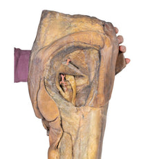
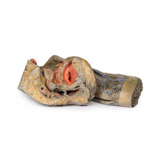
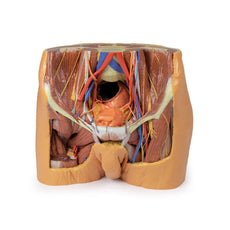
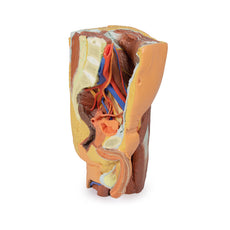
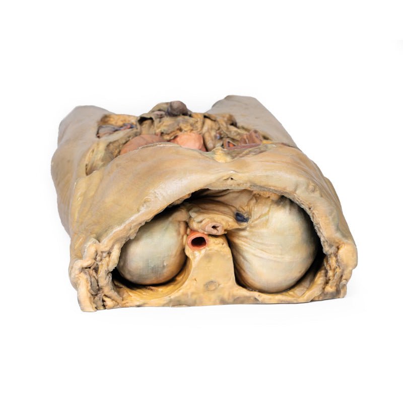
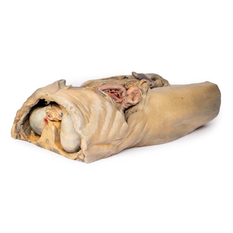
The inguinal region and perineum
A distinctive and unique feature of this model is the dissection of
simultaneous direct and indirect hernias preserved on the right and left sides, respectively. While most of the
anterior abdominal wall has bee removed, the inferior epigastric arteries (and accompanying veins) have been
retained to allow for interpretation of the herniations. On the right side, a distinct outpouching of the
parietal peritoneum has formed medial relative to the inferior epigastric artery, representing an indirect
herniation event. On the left side, the hernia sac extends laterally relative to the inferior epigastric artery
and into the opened spermatic cord, with continuity of the epiploic appendage from the sigmoid colon into the
sac.
The skin over the perineum has been removed in order to demonstrate both the structure of the penis
(with both the corpus spongiosum and corpora cavernosa contrasted) and the position of the testes and spermatic
cords relative to the anterior abdominal wall. On the right side, which in this individual is impacted by a
direct hernia, the spermatic cord has been left undissected allowing for an appreciation of the external
spermatic fascia from the inguinal region through to the testis. On the left side, the spermatic cord has been
opened and is dominated by the enlarged and varicose testicular vein (reflecting the impact of the indirect
hernia exposed within the cord) just superior to the epididymis and exposed tunica albuginea of the testis.
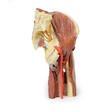
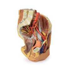
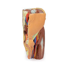
The thigh
Anterior dissections into the femoral triangle region have been undertaken to both thighs with
varying preservation of contents. On the right side the femoral sheath has been removed to expose the femoral
artery, vein and the deep inguinal lymph nodes. The femoral artery has been sectioned with a portion removed to
expose the origin of the profunda femoris and to better appreciate the draining of the great saphenous vein into
the femoral vein. Just lateral to these structures the very terminal component of the femoral nerve is visible.
On the left side a slightly larger dissection window has been opened to expose more of the underlying anterior
and medial thigh compartment muscles, from the sartorius and iliopsoas laterally to the pectineus and adductor
longus medially. The femoral artery has been preserved, with a well-preserved superficial circumflex iliac
artery and the origin of the profunda femoris visible adjacent to the femoral nerve.
The model terminates at
the level of the mid-thigh, and while not a primary focus of the model the spatial organisation of structures in
the cross-section can be seen. This includes the anteriorly positioned femoral diaphysis with tightly-packed
anterior compartment muscles and the passage of the femoral artery and vein in the subsartorial canal.
Handling Guidelines for 3D Printed Models
GTSimulators by Global Technologies
Erler Zimmer Authorized Dealer
These items normal warranty are two years, however the warranty doesn’t cover “wear and tear”. The manufacturer does have 100% quality control on these models.
The models are very detailed and delicate. With normal production machines you cannot realize such details like shown in these models.
The printer used is a color-plastic printer. This is the most suitable printer for these models.
The plastic material is already the best and most suitable material for these prints. (The other option would be a kind of gypsum, but this is way more fragile. You even cannot get them out of the printer without breaking them).The huge advantage of the prints is that they are very realistic as the data is coming from real human specimen. Nothing is shaped or stylized.
The users have to handle these prints with utmost care. They are not made for touching or bending any thin nerves, arteries, vessels etc. The 3D printed models should sit on a table and just rotated at the table.
Related Products


$3,141.00
$3,490.00
Free shipping
3D Printed Lower Limb - deep dissection of a left pelvis and thigh
Item # MP1813


$2,706.00
$3,007.00
Free shipping
3D Printed Female right pelvis superficial and deep structures
Item # MP1783


by — Item # MP1130
3D Printed Abdomen with Bilateral Hernias
$11,589.00
$12,877.00
Add to Cart
Add to Quote