Your shopping cart is empty.
3D Printed Female Pelvis Deep Dissection


 Handling Guidelines for 3D Printed Models
GTSimulators by Global Technologies
Erler Zimmer Authorized Dealer


3D Printed Female Pelvis Deep Dissection
Item # MP1141
$7,095.00
$7,884.00
You save $789.00
Need an estimate?
Click Add To Quote

Features & Specifications
-
 by
by
A trusted GT partner -
FREE Shipping
U.S. Contiguous States Only -
 3D Printed Model
from a real specimen -
 Gov't pricing
Available upon request
 by
by 
Frequently Bought Together

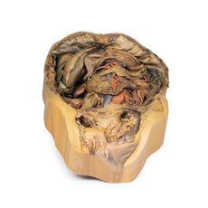
3D Printed Female Pelvis Deep Dissection
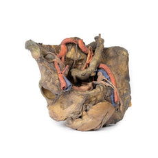
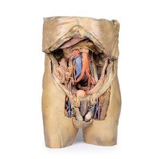
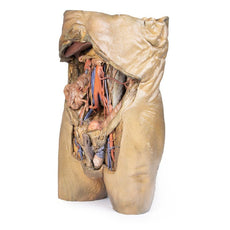
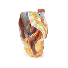
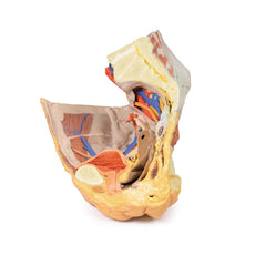
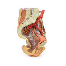
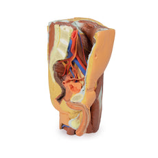
This 3D model presents a deep dissection and isolation of the pelvis from surrounding regions, particularly
demonstrating visceral and neurovascular structures relative to deep ligaments and osseous features.
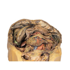
Within the false pelvis, the sigmoid colon descends on the left side of the specimen to the rectum, passing
superficially across the pelvic brim and the passage of the common and external iliac artery and vein. Adjacent
to the sigmoid colon are parts of the sigmoid arteries and superior rectal artery, resting superficial to the
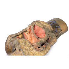
common iliac vessels and near the descending ureter. Anterior in the true pelvis is the collapsed urinary
bladder, and between the bladder and rectum rests the uterus. The organ is partially covered in the broad
ligament, with both the suspensory ligament of the ovary and round ligament have been separated and pulled away
from the peritoneum on both sides to expose surrounding blood vessels. While the ovarian ligaments, round
ligaments, uterine tubes and ovaries are trapped within the peritoneal fold of the broad ligament, the reduction
in ovary size (common with advanced age) has rendered these indistinguishable in the model.
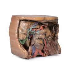
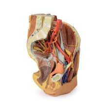
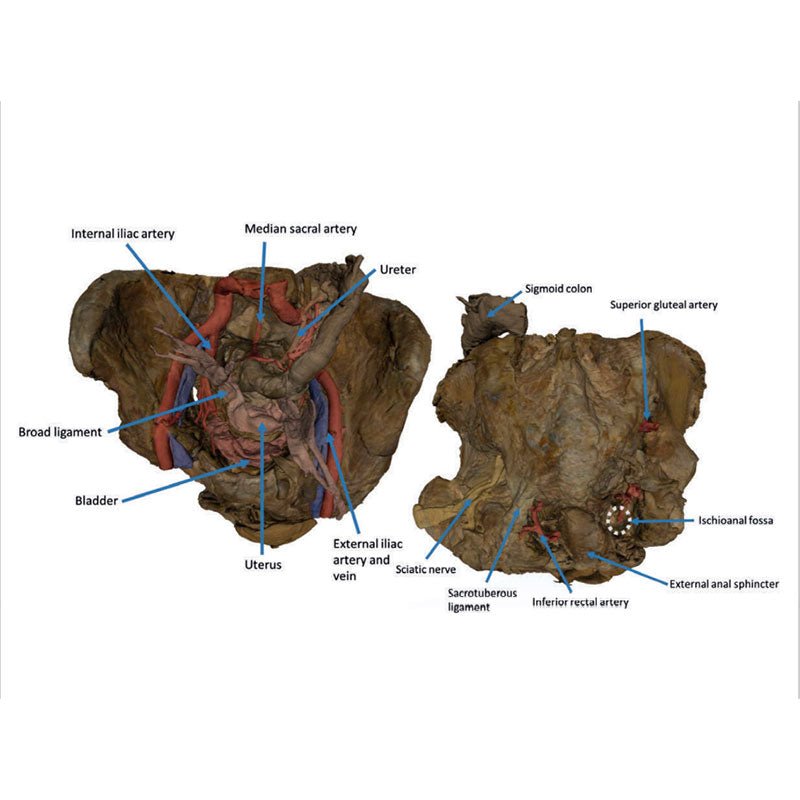
Lateral to these organs, branches of the internal iliac artery can be identified – as well as a retained median
sacral artery in the midline between the two common iliac arteries. On the left side only the uterine artery can
be seen laterally. On the right side, the obturator, superior vesical, and uterine arteries can be observed. In
addition, the origins of the inferior epigastric artery and vein can be seen arising from the external iliac
vessels just prior to exiting the inferior abdominal cavity.
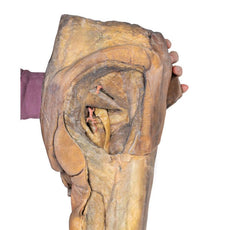
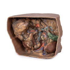
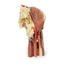
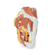
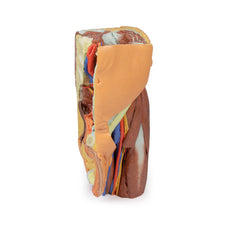
On the right side of the preserved pelvis, the entire femur and thigh musculature has been removed to
demonstrate the obturator membrane, the articular cartilage of the acetabulum and the transverse ligament of the
acetabulum. Posteriorly the entire gluteal region has been dissected to expose the superior gluteal foramen and
the origin of the superior gluteal artery. The sacrotuberous ligament has been removed to demonstrate the
sacrospinous ligament, with some branches of the inferior rectal artery retained within the exposed ischoanal
fossa.
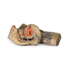
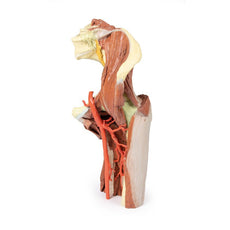
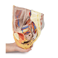
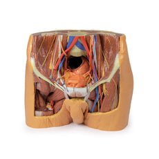
On the left side of the preserved pelvis the sciatic nerve has been maintained within the greater sciatic
foramen, as has the sacrotuberous ligament. The ischioanal fossa mirrors that of the right side, where branches
of the inferior rectal artery have been retained relative to the fibres of the pelvic diaphragm, and the
integration of the external anal sphincter on the projecting external rectal surface.
Handling Guidelines for 3D Printed Models
GTSimulators by Global Technologies
Erler Zimmer Authorized Dealer
These items normal warranty are two years, however the warranty doesn’t cover “wear and tear”. The manufacturer does have 100% quality control on these models.
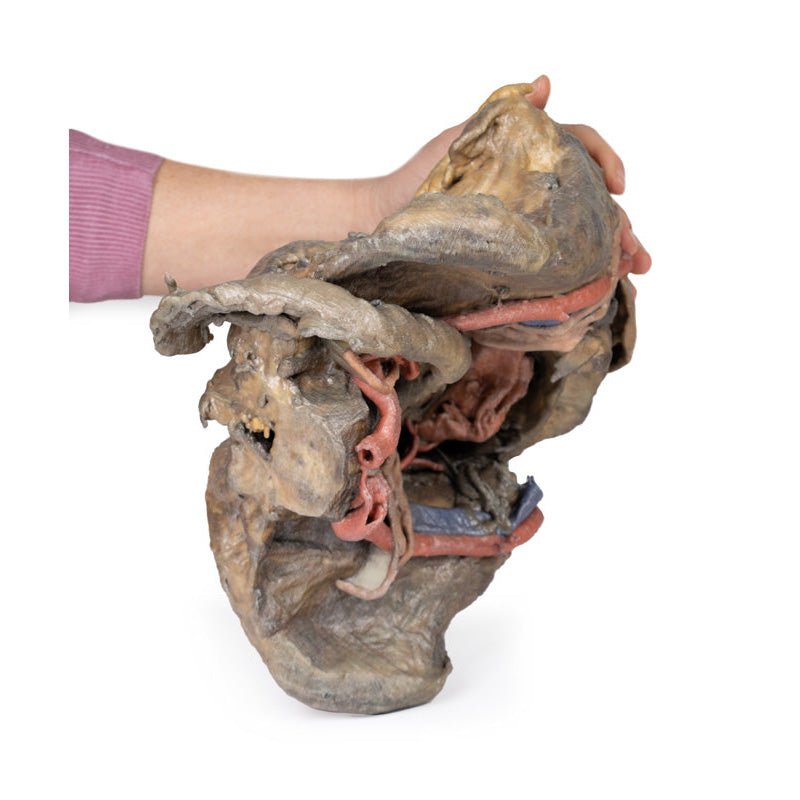
The models are very detailed and delicate. With normal production machines you cannot realize such details like shown in these models.
The printer used is a color-plastic printer. This is the most suitable printer for these models.
The plastic material is already the best and most suitable material for these prints. (The other option would be a kind of gypsum, but this is way more fragile. You even cannot get them out of the printer without breaking them).The huge advantage of the prints is that they are very realistic as the data is coming from real human specimen. Nothing is shaped or stylized.
The users have to handle these prints with utmost care. They are not made for touching or bending any thin nerves, arteries, vessels etc. The 3D printed models should sit on a table and just rotated at the table.
Related Products


$2,992.00
$3,361.00
Free shipping
3D Printed Lower Limb - deep dissection of a left pelvis and thigh
Item # MP1813


$2,578.00
$2,896.00
Free shipping
3D Printed Female right pelvis superficial and deep structures
Item # MP1783


by — Item # MP1141
3D Printed Female Pelvis Deep Dissection
$7,095.00
$7,884.00
Add to Cart
Add to Quote