

Your shopping cart is empty.
3D Printed Flexed knee joint deep dissection Model
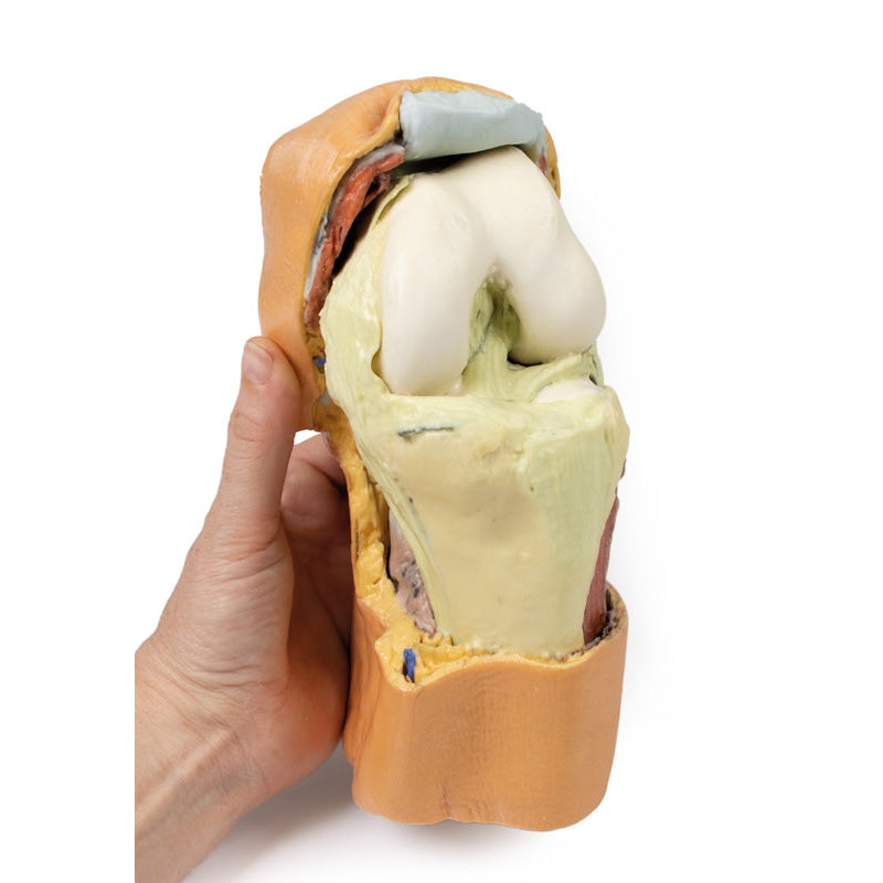

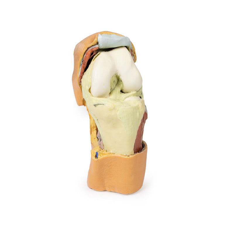
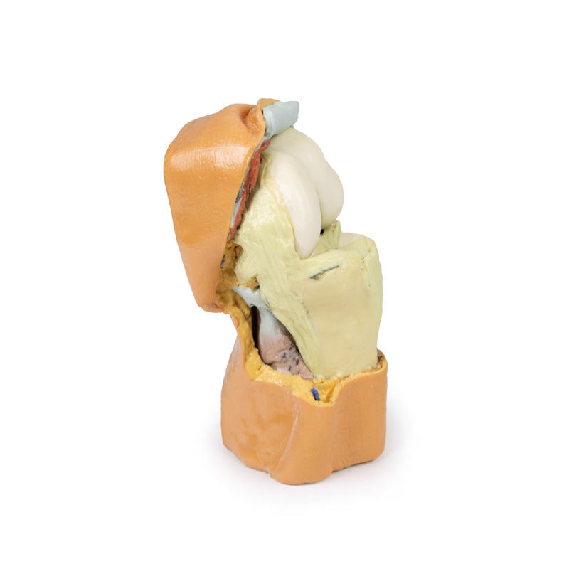
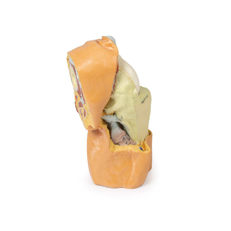
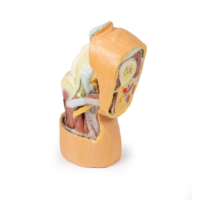
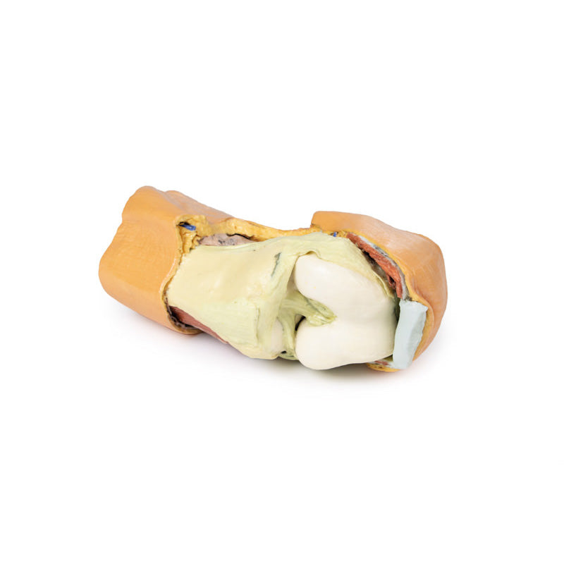
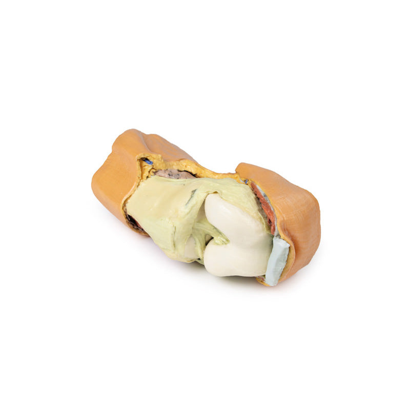
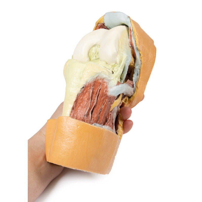
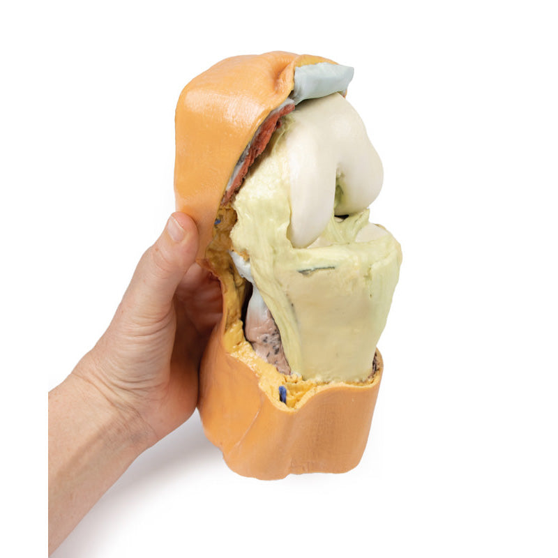
Anteriorly, the skin, subcutaneous tissue and patella have been removed, with only remnant portions of the tendon of the quadriceps femoris and patellar ligament retained. With the joint capsule opened, the anterior and posterior cruciate ligaments and the menisci are visible positioned between the femoral condyles and tibial plateau. On the medial aspect, the tibial (medial) collateral ligament passes just anterior to the insertion of the semitendinosus of the pes anserinus (the sartorius and gracilis tendons are sectioned), which in turn is just anterior to the posterior compartment musculature (covered by crural fascia). On the lateral aspect, the fibular (lateral) collateral ligament is preserved, and the anterior crural musculature is exposed. Passing from the thigh are the inserting tendon of the biceps femoris onto the head of the fibula, as well as the common peroneal nerve.
Download Handling Guidelines for 3D Printed Models
GTSimulators by Global Technologies
Erler Zimmer Authorized Dealer

8.0 lb


3D Printed Flexed knee joint deep dissection Model
Item # MP1807
$1,331.00
$1,479.00
You save $148.00
In stock
Need an estimate?
Click Add To Quote

Features & Specifications
-
 by
by
A trusted GT partner -
 3D Printed Model
from a real specimen -
 Gov't pricing
Available upon request
 by
by 
Frequently Bought Together

3D Printed Flexed knee joint deep dissection Model
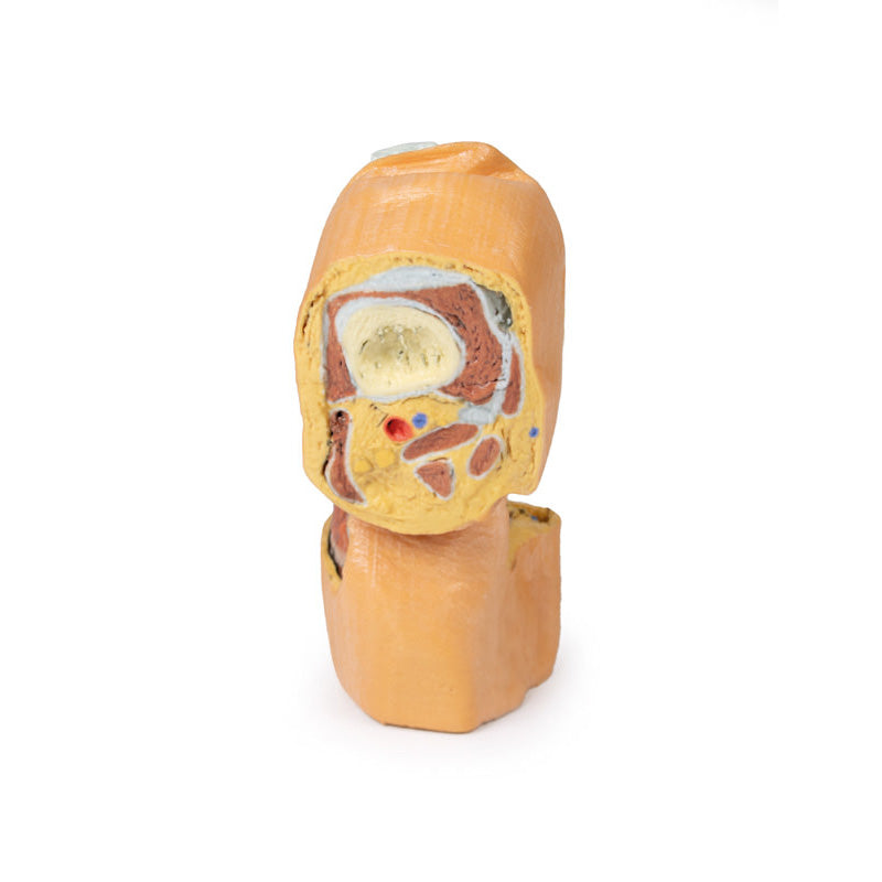
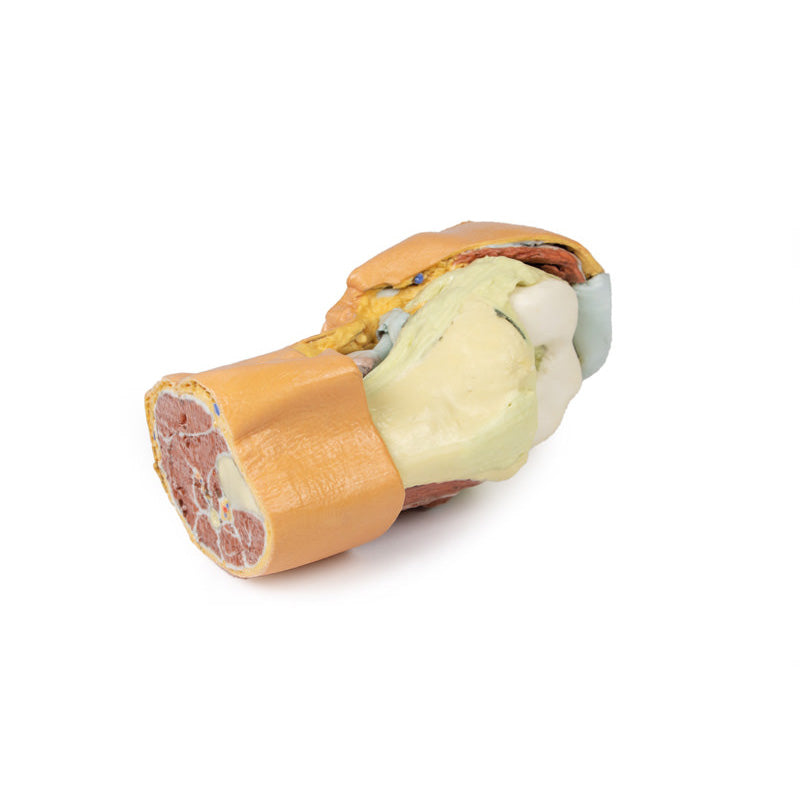
This 3D printed specimen displays a deep dissection of a left knee joint with the internal joint capsule structures relative to superficial tissues in a flexed position. The proximal cross-section through the distal thigh captures a small portion of the quadriceps femoris and sartorius anteriorly (with the thickened connective tissue of the iliotibial tract), the fat-filled popliteal fossa (with the popliteal vessels, tibial and common peroneal nerves), and the termination of the medial (adductor magnus tendon, gracilis) and posterior thigh muscles (biceps femoris, semitendinosus, semimembranosus) posteriorly. The distal cross-section through the leg preserves the most proximal portion of the muscles of the anterior, lateral and posterior compartments. Also visible in the section are the associated neurovascular structures: the anterior tibial artery, vein and deep peroneal nerve; the posterior tibial artery, vein and tibial nerve; and the fibular artery and vein.Anteriorly, the skin, subcutaneous tissue and patella have been removed, with only remnant portions of the tendon of the quadriceps femoris and patellar ligament retained. With the joint capsule opened, the anterior and posterior cruciate ligaments and the menisci are visible positioned between the femoral condyles and tibial plateau. On the medial aspect, the tibial (medial) collateral ligament passes just anterior to the insertion of the semitendinosus of the pes anserinus (the sartorius and gracilis tendons are sectioned), which in turn is just anterior to the posterior compartment musculature (covered by crural fascia). On the lateral aspect, the fibular (lateral) collateral ligament is preserved, and the anterior crural musculature is exposed. Passing from the thigh are the inserting tendon of the biceps femoris onto the head of the fibula, as well as the common peroneal nerve.
Download Handling Guidelines for 3D Printed Models
GTSimulators by Global Technologies
Erler Zimmer Authorized Dealer
These items normal warranty are two years, however the warranty doesn’t cover “wear and tear”. The manufacturer does have 100% quality control on these models.
The models are very detailed and delicate. With normal production machines you cannot realize such details like shown in these models.
The printer used is a color-plastic printer. This is the most suitable printer for these models.
The plastic material is already the best and most suitable material for these prints. (The other option would be a kind of gypsum, but this is way more fragile. You even cannot get them out of the printer without breaking them).The huge advantage of the prints is that they are very realistic as the data is coming from real human specimen. Nothing is shaped or stylized.
The users have to handle these prints with utmost care. They are not made for touching or bending any thin nerves, arteries, vessels etc. The 3D printed models should sit on a table and just rotated at the table.
Related Products


by — Item # MP1807
3D Printed Flexed knee joint deep dissection Model
$1,331.00
$1,479.00
Add to Cart
Add to Quote
