

Your shopping cart is empty.
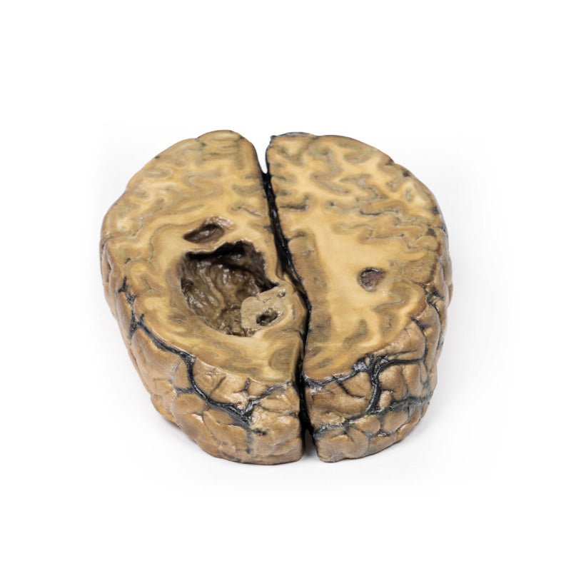
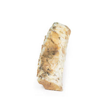
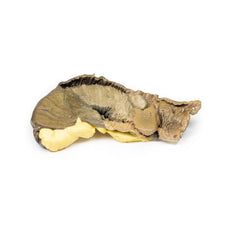
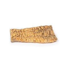
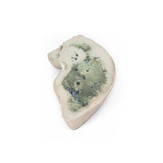
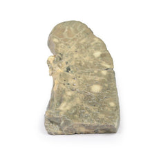
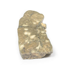
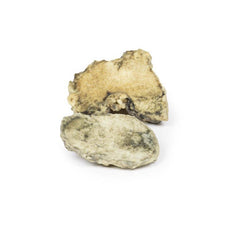
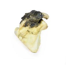
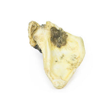
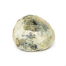
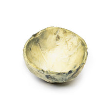
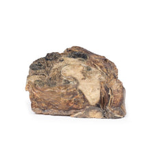
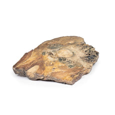
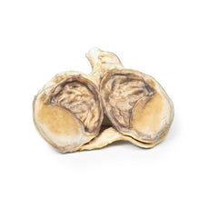
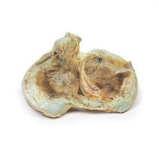
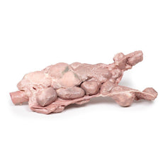
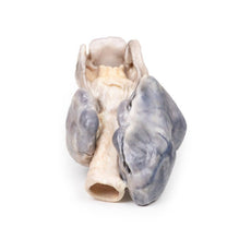
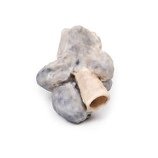
3D Printed Metastatic Carcinoma in the Brain

 Handling Guidelines for 3D Printed Models
GTSimulators by Global Technologies
Erler Zimmer Authorized Dealer


0.0 lb


3D Printed Metastatic Carcinoma in the Brain
Item # MP2019
$866.00
$963.00
You save $97.00
Need an estimate?
Click Add To Quote

Features & Specifications
-
 by
by
A trusted GT partner -
FREE Shipping
U.S. Contiguous States Only -
 3D Printed Model
from a real specimen -
 Gov't pricing
Available upon request
 by
by 
Frequently Bought Together

3D Printed Metastatic Carcinoma in the Brain
Clinical History (pre access to CT and MRI imaging)
This 51-year old woman had surgery for breast carcinoma 2
years before presentation. Her main complaint was left-sided ataxia for the 2 weeks prior, and this had been
preceded by a fainting attack followed by left-sided weakness. Examination revealed a left spastic paresis.
There was doubt as to the diagnosis because the rapidity of onset suggested a vascular lesion. She was
discharged from hospital but six weeks after her initial presentation she was re-admitted with left-sided
fitting. Lumber puncture and re-examination were not informative. EEG showed a right anterior temporal
abnormality. Angiography confirmed the presence of a large space-occupying lesion in the right cerebrum. On the
ward, there was a steady deterioration of the patient’s condition, and ultimately death.
Pathology
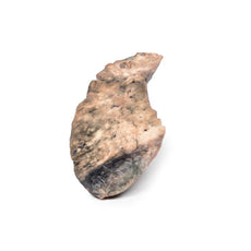
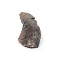
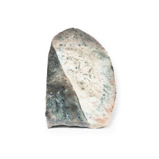
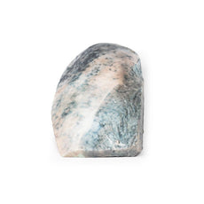
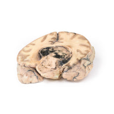
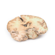
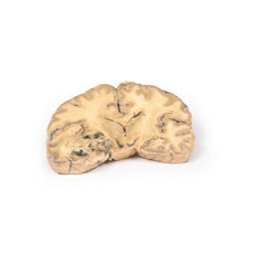
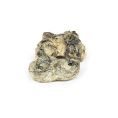
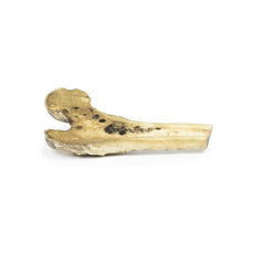
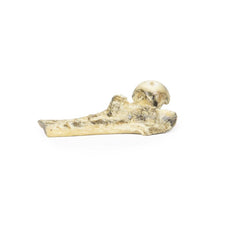
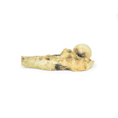
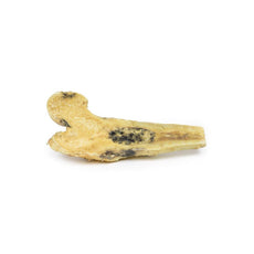
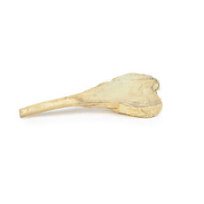
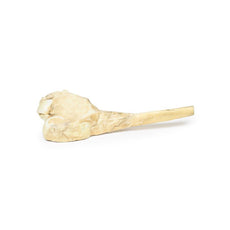
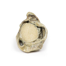
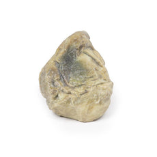
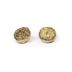
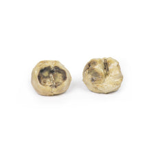
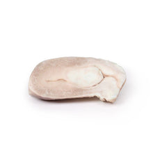
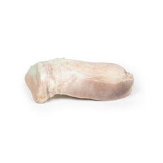
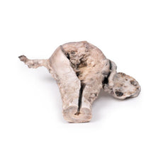
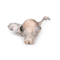
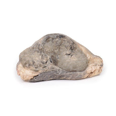
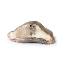
The specimen is the cerebrum sliced horizontally. On the superior view, the right hemisphere is
clearly enlarged, particularly in the parietal region where the gyrae are widened and 3 cystic tumours are
evident. The largest, 5 cm in diameter, is in the right parietal region. A smaller tumour, 2 x 1.5 cm in
diameter, is seen close to the posterior margin of the largest tumour. A third one, 1.5 cm in diameter, is
present in the left parietal region. The tumours have mainly involved white matter. The wall of each lesion is
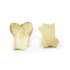
composed of shaggy friable greyish tissue. At necropsy, there was ulceration of the largest tumour into the
right lateral ventricle (seen more clearly when the inferior surface is examined). Sub-falcine herniation was
also seen, as is displacement of the basal ganglia and internal capsule. Histological examination revealed
metastatic carcinoma in the viable areas. Other metastases were found in the liver and bone. Histology of a
liver metastasis was consistent with origin from a primary carcinoma of breast.
Handling Guidelines for 3D Printed Models
GTSimulators by Global Technologies
Erler Zimmer Authorized Dealer
These items normal warranty are two years, however the warranty doesn’t cover “wear and tear”. The manufacturer does have 100% quality control on these models.
The models are very detailed and delicate. With normal production machines you cannot realize such details like shown in these models.
The printer used is a color-plastic printer. This is the most suitable printer for these models.
The plastic material is already the best and most suitable material for these prints. (The other option would be a kind of gypsum, but this is way more fragile. You even cannot get them out of the printer without breaking them).The huge advantage of the prints is that they are very realistic as the data is coming from real human specimen. Nothing is shaped or stylized.
The users have to handle these prints with utmost care. They are not made for touching or bending any thin nerves, arteries, vessels etc. The 3D printed models should sit on a table and just rotated at the table.
Related Products


by — Item # MP2019
3D Printed Metastatic Carcinoma in the Brain
$866.00
$963.00
Add to Cart
Add to Quote
