Your shopping cart is empty.
3D Printed Temporal Bone Model, Set of 3
Part 1 Skull Preparation
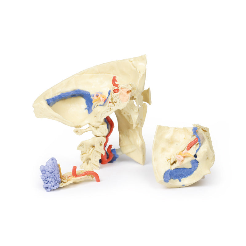
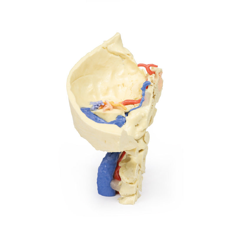
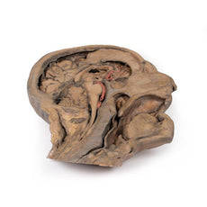
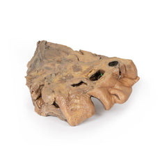
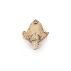
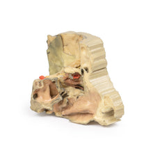
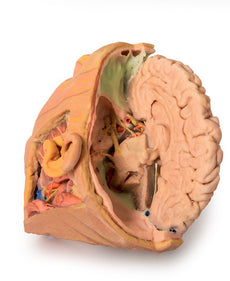
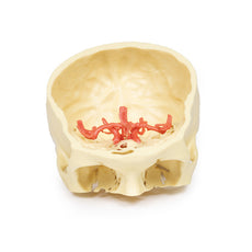
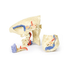
The specimen has been trimmed to reveal the posterior quadrant of the left side of the skull including the posterior (cerebellar part only) and middle cranial fossa. The model shows the location of the temporal bone and its relationship with the adjoining sphenoid, parietal and occipital bones. The superior aspect of the petrous part of the temporal bone including the tegmen tympani has been removed to reveal its detailed internal architecture, and structure associated with the auditory and vestibular apparatus. The middle ear (coloured orange) is revealed to show the tympanum, along with the aditus, antrum (laterally), and the ‘bone’ part of the pharangotympanic tube and the bony canal of the tensor tympani muscle (medially). Collectively, these form a direct anterior-posterior passage between the nasopharynx and the mastoid air cells (coloured blue). The anatomical position of the incus relative to the tympanic membrane can be seen via the external auditory meatus. The bony labyrinth of the vestibular apparatus of the inner ear (green) is seen juxtaposed with the middle ear. The orthogonal arrangement of the anterior, lateral, and posterior semicircular canals and the spiral organisation of the cochlea can be clearly identified. The passage of the facial nerve (CN VII) through the petrous part of the temporal bone and its intimate spatial relationship with the auditory and vestibular apparatus is shown in yellow. Proximad, the nerve courses in an anterolateral direction before descending distally to emerge from the bone via the stylomastoid foramen located between the mastoid and styloid processes. The condyle of the mandible can be seen in the mandibular fossa at the origin of the zygomatic process of squamous part of the temporal bone. The temporomandibular joint has had the capsule removed to reveal the articular disc of the joint (indicated by a blue/grey colouration). The anterior aspect of the mastoid process has been transected to show the extensive nature of the mastoid air cells. The cervical part of the internal carotid artery can be seen ascending to enter into the carotid canal within the petrous part of the temporal bone. Its anteromedial course can be seen within the exposed aspect of the bone, and its s-shaped continuance within the cavernous sinus of the sphenoid bone, and its emergence into the neurocranium. The model also shows the transverse dural venous sinus, its continuation into the sigmoidal sinus (located on the posterior internal aspect of the squamous part of the temporal bone), and passage through the jugular foramen to form the internal jugular vein. The inferior petrosal sinus is also seen leading into the jugular foramen. The model also shows the foramen magnum and first three cervical vertebrae cut in parasagittal section. Note the sphenoid sinus located axially within the base of the sphenoid bone (coloured blue)
Part 2 The Petrous Part Of The Temporal Bone
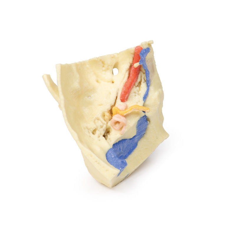
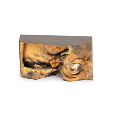
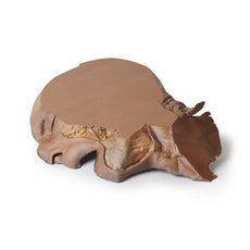
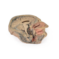
This model is derived from the overall skull preparation and has been enlarged (x3) to further illustrate the detailed internal architecture of the petrous part of the temporal bone and the auditory and vestibular apparatus. As in Part 1 internal casts of the bony labyrinth of the inner ear, mastoid air cells, and the bony canal of the internal carotid artery are used to aid comprehension of this complex and important region. The bony ossicles of the middle ear (incus, malleus and stapes) are shown within the middle ear cavity, and the bony prominence of the lateral semicircular canal of the vestibular apparatus can be seen protruding into the middle ear. The connection from the tympanum or middle ear to the mastoid aid cells (Blue) via the aditus and antrum is visible. The model also shows the facial nerve entering the internal acoustic meatus on the anterior aspect of the petrous bone. The chorda tympani is seen branching from the facial nerve in its descending portion in the facial canal. This small nerve, which carries parasympathetic fibres and taste fibres to the anterior 2/3 of the tongue, can be seen passing through the tympanic cavity between the incus and malleus. The bone canal of the tensor tympani muscle can be seen extending away from the tympanic cavity of the middle ear.
Part 3 The Auditory And Vestibular Apparatus
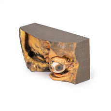
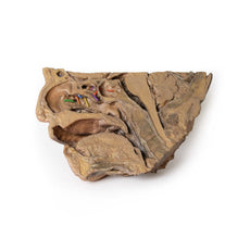
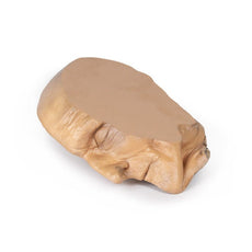
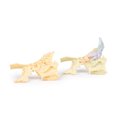
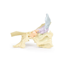
As in Part 2, this model has been enlarged (x3) to highlight the detailed internal architecture of the auditory and vestibular apparatus and its relationship to anatomical features of otological importance. The petrous part of the temporal bone and tegmen tympani have been removed to expose the tympanum, aditus and antrum of the tympanic cavity of the middle ear. The model shows the direct connection between nasopharanx and mastoid air cells via the bony canal of the pharangotympanic tube. The ossicles of the middle ear are seen within the tympanum or middle ear. The model also shows the bone labyrinth of the vestibular apparatus of the inner ear, and the tympanic prominence of the lateral semicircular canal can be seen within the tympanum of the middle ear. The passage of the facial (CN VII) and vestibulocochlear (CN VIII) nerves through the petrous part of the temporal bone, and their intimate spatial relationship with the auditory and vestibular apparatus is shown in yellow. The cochlear nerve is seen entering the cochlear region of the bony labyrinth of the inner ear. The genu and geniculate ganglion of the facial nerve are shown immediately before the facial nerve descends within the facial canal, to emerge via the stylomastoid foramen located between the mastoid and styloid processes. The chorda tympani is seen within the tympanum of the middle ear, passing between the incus and malleus. The emergence of the chorda tympani is shown from the petrotympanic fissure, located medial to the mandibular fossa of the temporal bone.
Download Handling Guidelines for 3D Printed Models
GTSimulators by Global Technologies
Erler Zimmer Authorized Dealer

3D Printed Temporal Bone Model, Set of 3
Item # MP1620
$2,083.00
$2,339.00
You save $256.00
Need an estimate?
Click Add To Quote

Features & Specifications
-
by
A trusted GT partner -
FREE Shipping
U.S. Contiguous States Only -
3D Printed Model
from a real specimen -
Gov't pricing
Available upon request
Frequently Bought Together
3D Printed Temporal Bone Model, Set of 3
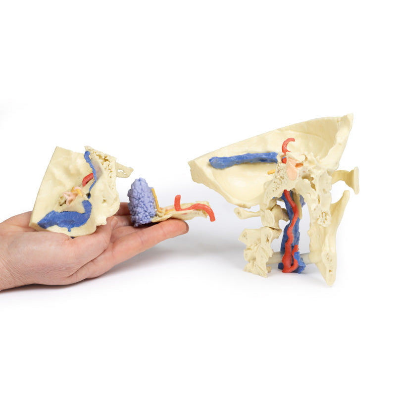
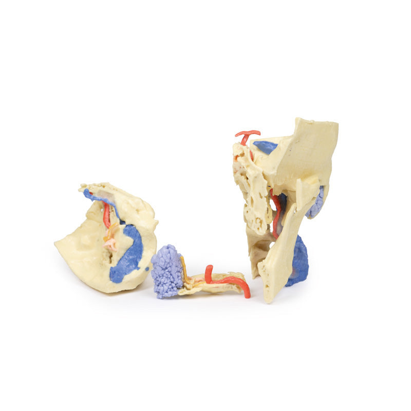
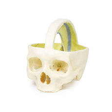
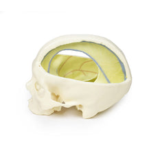
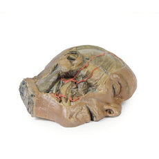
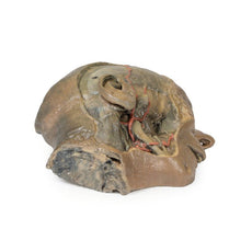
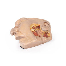
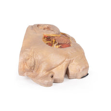
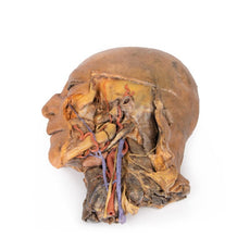
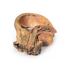
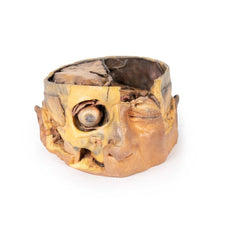
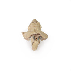
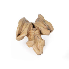
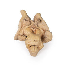
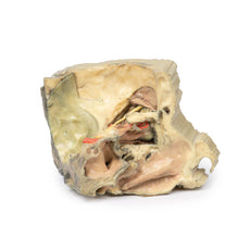
This 3 part 3D printed model derived from CT data highlights the complex anatomy of the temporal bone including bone ossicles, canals, chambers, foramina and air spaces. In addition, the spatial relations between temporal bone and other structures of otological importance, i.e. carotid artery, dural venous sinuses, related nerves and the dura mater are indicated. Internal casts (endocasts) of the bony chambers and canals have been created to aid visualisation of the internal anatomy of the temporal bonePart 1 Skull Preparation
The specimen has been trimmed to reveal the posterior quadrant of the left side of the skull including the posterior (cerebellar part only) and middle cranial fossa. The model shows the location of the temporal bone and its relationship with the adjoining sphenoid, parietal and occipital bones. The superior aspect of the petrous part of the temporal bone including the tegmen tympani has been removed to reveal its detailed internal architecture, and structure associated with the auditory and vestibular apparatus. The middle ear (coloured orange) is revealed to show the tympanum, along with the aditus, antrum (laterally), and the ‘bone’ part of the pharangotympanic tube and the bony canal of the tensor tympani muscle (medially). Collectively, these form a direct anterior-posterior passage between the nasopharynx and the mastoid air cells (coloured blue). The anatomical position of the incus relative to the tympanic membrane can be seen via the external auditory meatus. The bony labyrinth of the vestibular apparatus of the inner ear (green) is seen juxtaposed with the middle ear. The orthogonal arrangement of the anterior, lateral, and posterior semicircular canals and the spiral organisation of the cochlea can be clearly identified. The passage of the facial nerve (CN VII) through the petrous part of the temporal bone and its intimate spatial relationship with the auditory and vestibular apparatus is shown in yellow. Proximad, the nerve courses in an anterolateral direction before descending distally to emerge from the bone via the stylomastoid foramen located between the mastoid and styloid processes. The condyle of the mandible can be seen in the mandibular fossa at the origin of the zygomatic process of squamous part of the temporal bone. The temporomandibular joint has had the capsule removed to reveal the articular disc of the joint (indicated by a blue/grey colouration). The anterior aspect of the mastoid process has been transected to show the extensive nature of the mastoid air cells. The cervical part of the internal carotid artery can be seen ascending to enter into the carotid canal within the petrous part of the temporal bone. Its anteromedial course can be seen within the exposed aspect of the bone, and its s-shaped continuance within the cavernous sinus of the sphenoid bone, and its emergence into the neurocranium. The model also shows the transverse dural venous sinus, its continuation into the sigmoidal sinus (located on the posterior internal aspect of the squamous part of the temporal bone), and passage through the jugular foramen to form the internal jugular vein. The inferior petrosal sinus is also seen leading into the jugular foramen. The model also shows the foramen magnum and first three cervical vertebrae cut in parasagittal section. Note the sphenoid sinus located axially within the base of the sphenoid bone (coloured blue)
Part 2 The Petrous Part Of The Temporal Bone
This model is derived from the overall skull preparation and has been enlarged (x3) to further illustrate the detailed internal architecture of the petrous part of the temporal bone and the auditory and vestibular apparatus. As in Part 1 internal casts of the bony labyrinth of the inner ear, mastoid air cells, and the bony canal of the internal carotid artery are used to aid comprehension of this complex and important region. The bony ossicles of the middle ear (incus, malleus and stapes) are shown within the middle ear cavity, and the bony prominence of the lateral semicircular canal of the vestibular apparatus can be seen protruding into the middle ear. The connection from the tympanum or middle ear to the mastoid aid cells (Blue) via the aditus and antrum is visible. The model also shows the facial nerve entering the internal acoustic meatus on the anterior aspect of the petrous bone. The chorda tympani is seen branching from the facial nerve in its descending portion in the facial canal. This small nerve, which carries parasympathetic fibres and taste fibres to the anterior 2/3 of the tongue, can be seen passing through the tympanic cavity between the incus and malleus. The bone canal of the tensor tympani muscle can be seen extending away from the tympanic cavity of the middle ear.
Part 3 The Auditory And Vestibular Apparatus
As in Part 2, this model has been enlarged (x3) to highlight the detailed internal architecture of the auditory and vestibular apparatus and its relationship to anatomical features of otological importance. The petrous part of the temporal bone and tegmen tympani have been removed to expose the tympanum, aditus and antrum of the tympanic cavity of the middle ear. The model shows the direct connection between nasopharanx and mastoid air cells via the bony canal of the pharangotympanic tube. The ossicles of the middle ear are seen within the tympanum or middle ear. The model also shows the bone labyrinth of the vestibular apparatus of the inner ear, and the tympanic prominence of the lateral semicircular canal can be seen within the tympanum of the middle ear. The passage of the facial (CN VII) and vestibulocochlear (CN VIII) nerves through the petrous part of the temporal bone, and their intimate spatial relationship with the auditory and vestibular apparatus is shown in yellow. The cochlear nerve is seen entering the cochlear region of the bony labyrinth of the inner ear. The genu and geniculate ganglion of the facial nerve are shown immediately before the facial nerve descends within the facial canal, to emerge via the stylomastoid foramen located between the mastoid and styloid processes. The chorda tympani is seen within the tympanum of the middle ear, passing between the incus and malleus. The emergence of the chorda tympani is shown from the petrotympanic fissure, located medial to the mandibular fossa of the temporal bone.
Download Handling Guidelines for 3D Printed Models
GTSimulators by Global Technologies
Erler Zimmer Authorized Dealer
These items normal warranty are two years, however the warranty doesn’t cover “wear and tear”. The manufacturer does have 100% quality control on these models.
The models are very detailed and delicate. With normal production machines you cannot realize such details like shown in these models.
The printer used is a color-plastic printer. This is the most suitable printer for these models.
The plastic material is already the best and most suitable material for these prints. (The other option would be a kind of gypsum, but this is way more fragile. You even cannot get them out of the printer without breaking them).The huge advantage of the prints is that they are very realistic as the data is coming from real human specimen. Nothing is shaped or stylized.
The users have to handle these prints with utmost care. They are not made for touching or bending any thin nerves, arteries, vessels etc. The 3D printed models should sit on a table and just rotated at the table.

by — Item # MP1620
3D Printed Temporal Bone Model, Set of 3
$2,083.00
$2,339.00
Add to Cart
Add to Quote


